File:Mouse spermiogenesis 01.jpg

Original file (1,200 × 299 pixels, file size: 48 KB, MIME type: image/jpeg)
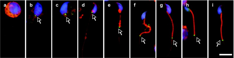
Spatiotemporal progression of annulus during mouse spermiogenesis.
The annulus (arrows) indicated by the SEPT4 antibody was first seen at the neck region of step 9 spermatids (b), not in step 8 spermatids (a). It stayed there until early step 15 (c-d), and after that it migrated to its distal position followed by the formation of mitochondrial sheath (e-h). It still existed in the mature spermatiozoon coonecting the midpiece and the principal piece of the tail (i).
Green, SEPT4; Red, MitoTracker; Blue, DAPI.
The scale bar represents 10 μm.
1471-213X-9-23-2-l.jpg
http://www.biomedcentral.com/1471-213X/9/23#B1
Guan et al. BMC Developmental Biology 2009 9:23 doi:10.1186/1471-213X-9-23
Spatiotemporal association of DNAJB13 with the annulus during mouse sperm flagellum development. Guan J, Kinoshita M, Yuan L. BMC Dev Biol. 2009 Mar 19;9:23. PMID: 19298648
© 2009 Guan et al; licensee BioMed Central Ltd. This is an Open Access article distributed under the terms of the Creative Commons Attribution License (http://creativecommons.org/licenses/by/2.0), which permits unrestricted use, distribution, and reproduction in any medium, provided the original work is properly cited.
File history
Click on a date/time to view the file as it appeared at that time.
| Date/Time | Thumbnail | Dimensions | User | Comment | |
|---|---|---|---|---|---|
| current | 13:35, 5 April 2010 | 1,200 × 299 (48 KB) | S8600021 (talk | contribs) | Spatiotemporal progression of annulus during mouse spermiogenesis. The annulus (arrows) indicated by the SEPT4 antibody was first seen at the neck region of step 9 spermatids (b), not in step 8 spermatids (a). It stayed there until early step 15 (c-d), |
You cannot overwrite this file.
File usage
The following page uses this file: