File:Mouse oocyte balbini body EM01.jpg

Original file (695 × 700 pixels, file size: 155 KB, MIME type: image/jpeg)
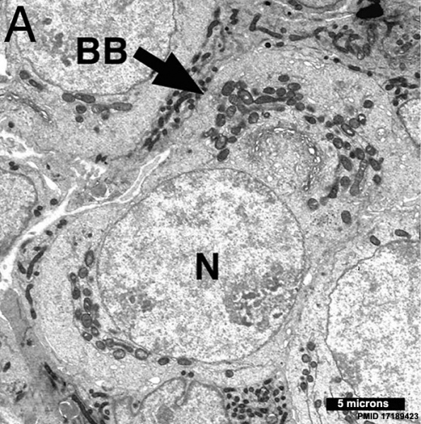
Mouse Oocyte Balbini Body
Electron micrograph of oocytes in neonatal ovary micrograph of an oocyte within a germline cyst from PND1 showing a well defined Balbiani body (arrow) with Golgi surrounded by mitochondria.
- Balbiani body (mitochondrial cloud) is a large organelle aggregate found in developing oocytes of many species.
- EM Links: All Images | balbini_body | A | B | C | D | E | F | Oocyte Development | Ovary Development | Mouse Development
Reference
Pepling ME, Wilhelm JE, O'Hara AL, Gephardt GW & Spradling AC. (2007). Mouse oocytes within germ cell cysts and primordial follicles contain a Balbiani body. Proc. Natl. Acad. Sci. U.S.A. , 104, 187-92. PMID: 17189423 DOI.
Copyright
Proceedings National Academy of Sciences (PNAS) Liberalization of PNAS copyright policy: Noncommercial use freely allowed Note original Author should be contacted for permission to reuse for Educational purposes. See also PNAS Author Rights and Permission FAQs
- Cozzarelli NR, Fulton KR, Sullenberger DM. Liberalization of PNAS copyright policy: noncommercial use freely allowed. Proc Natl Acad Sci U S A. 2004 Aug 24;101(34):12399. PMID15314225 "Our guiding principle is that, while PNAS retains copyright, anyone can make noncommercial use of work in PNAS without asking our permission, provided that the original source is cited."
Cite this page: Hill, M.A. (2024, April 20) Embryology Mouse oocyte balbini body EM01.jpg. Retrieved from https://embryology.med.unsw.edu.au/embryology/index.php/File:Mouse_oocyte_balbini_body_EM01.jpg
- © Dr Mark Hill 2024, UNSW Embryology ISBN: 978 0 7334 2609 4 - UNSW CRICOS Provider Code No. 00098G
File history
Click on a date/time to view the file as it appeared at that time.
| Date/Time | Thumbnail | Dimensions | User | Comment | |
|---|---|---|---|---|---|
| current | 13:43, 9 May 2013 | | 695 × 700 (155 KB) | Z8600021 (talk | contribs) | ==Mouse oocyte balbini body== Electron micrographs of oocytes in neonatal ovaries. (A) Micrograph of an oocyte within a germline cyst from PND1 showing a well defined Balbiani body (arrow) with Golgi surrounded by mitochondria. {{PNAS}} |
You cannot overwrite this file.
File usage
The following 2 pages use this file: