File:Mouse - palate MMP-25 expression.jpg

Original file (1,000 × 818 pixels, file size: 243 KB, MIME type: image/jpeg)
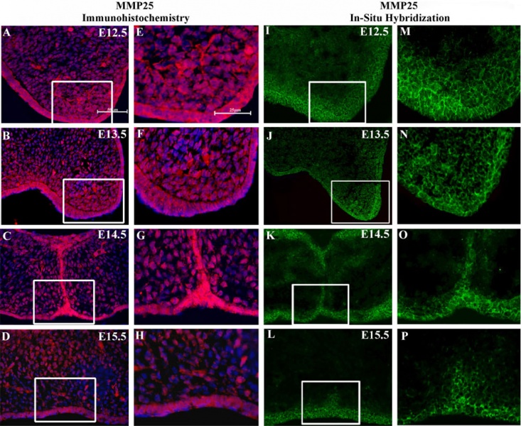
Mouse Palate MMP-25 Expression
Localization of matrix metalloproteinase-25 (MMP-25) protein and mRNA expression in the developing mouse palate.
- A-H - Immunofluorescent images of MMP-25 protein expression (red) colocalized with Hoechst nuclear staining (blue).
- I-P - In situ hybridization of MMP-25 mRNA expression (green).
Expression of MMP-25 appears stronger in the epithelium of the palate shelves than in the underlying mesenchyme. (E-H) Enhanced views of highlighted areas from A-D. (M,P) Enhanced views of highlighted areas from I-L.
For A-D and I-L, scale bar indicates 50 μm. For E-H and M-P, scale bar indicates 25 μm.
Reference
<pubmed>20809987</pubmed>| PMC2944159
Brown and Nazarali BMC Developmental Biology 2010 10:93 doi:10.1186/1471-213X-10-93
Copyright
© 2010 Brown and Nazarali; licensee BioMed Central Ltd. This is an Open Access article distributed under the terms of the Creative Commons Attribution License (http://creativecommons.org/licenses/by/2.0), which permits unrestricted use, distribution, and reproduction in any medium, provided the original work is properly cited.
Original File Name: Figure 2. 1471-213X-10-93-2-l.jpg http://www.biomedcentral.com/1471-213X/10/93/figure/F2
File history
Click on a date/time to view the file as it appeared at that time.
| Date/Time | Thumbnail | Dimensions | User | Comment | |
|---|---|---|---|---|---|
| current | 14:00, 2 November 2010 | | 1,000 × 818 (243 KB) | S8600021 (talk | contribs) | ==Mouse Palate MMP-25 Expression== Localization of matrix metalloproteinase-25 (MMP-25) protein and mRNA expression in the developing mouse palate. (A-H) Immunofluorescent images of MMP-25 protein expression (red) colocalized with Hoechst nuclear stain |
You cannot overwrite this file.
File usage
The following 2 pages use this file: