File:Mouse - Spry1 cleft palate.jpg

Original file (600 × 848 pixels, file size: 166 KB, MIME type: image/jpeg)
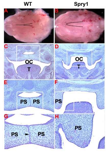
Spry1_Wnt1-Cre embryos exhibit cleft palate
(A, B) The developing mandible and tongue were removed from E14.5 embryos to show the developing medial epithelial seam of the palatal shelves in WT but not in Spry1;Wnt1-Cre (Spry1) embryos (dash line indicated), (C-H) cross sections of E16.5 embryo heads stained with hematoxylin and eosin.
(C, D) Low magnification shows the fused palate and the separated nasopharynx and oral cavity in WT control, but not in Spry1;Wnt1-Cre embryos (from original 10 ×), (E, F) high magnification from the boxed areas in C and D (from original 10 ×), (G, H) high magnification from the boxed areas of E and F
(from original 20 ×).
OC: oral cavity; PS: palatal shelves; T: tongue. Data are representative of four litters analyzed as this stage.
Original file name: Figure 3. 1471-213X-10-48-3.jpg
Reference
<pubmed>20459789</pubmed>| BMC Developmental Biology
© 2010 Yang et al; licensee BioMed Central Ltd.
This is an Open Access article distributed under the terms of the Creative Commons Attribution License (http://creativecommons.org/licenses/by/2.0), which permits unrestricted use, distribution, and reproduction in any medium, provided the original work is properly cited.
File history
Click on a date/time to view the file as it appeared at that time.
| Date/Time | Thumbnail | Dimensions | User | Comment | |
|---|---|---|---|---|---|
| current | 09:40, 28 January 2011 |  | 600 × 848 (166 KB) | S8600021 (talk | contribs) | ==Spry1_Wnt1-Cre embryos exhibit cleft palate== (A, B) The developing mandible and tongue were removed from E14.5 embryos to show the developing medial epithelial seam of the palatal shelves in WT but not in Spry1;Wnt1-Cre (Spry1) embryos (dash line indi |
You cannot overwrite this file.
File usage
The following 2 pages use this file: