File:Mouse- seminiferous tubule histology.jpg
Mouse-_seminiferous_tubule_histology.jpg (715 × 427 pixels, file size: 76 KB, MIME type: image/jpeg)
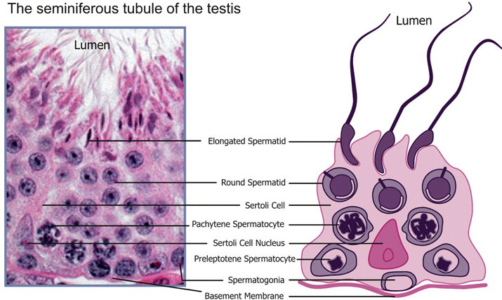
Mouse Seminiferous Tubule Histology
The seminiferous tubule of the testis (a cross-section): the Sertoli cells provide support and nutrients for the developing germ cells.
Sertoli cells also form the blood–testis barrier between adjacent Sertoli cells, which functionally divide the seminiferous epithelium into the basal and luminal compartments.
Spermatogonia, the self-renewing stem cells of the testis, are associated with the basement membrane of the tubule. As the germ cells develop, from spermatocytes to spermatids, they move progressively closer to the lumen of the tubule where they are released in a process known as spermiation.
Original File Name: Figure 1 http://www.ncbi.nlm.nih.gov/pmc/articles/PMC2816191/figure/DMP032F1/ (A extracted from full figure)
Reference
<pubmed>19758979</pubmed>| PMC2816191 | Hum Reprod Update.
From: Hum Reprod Update. 2010 Mar–Apr; 16(2): 205–224. Published online 2009 September 15. doi: 10.1093/humupd/dmp032.
This is an Open Access article distributed under the terms of the Creative Commons Attribution Non-Commercial License (http://creativecommons.org/licenses/by-nc/2.5/uk/) which permits unrestricted non-commercial use, distribution, and reproduction in any medium, provided the original work is properly cited.
File history
Click on a date/time to view the file as it appeared at that time.
| Date/Time | Thumbnail | Dimensions | User | Comment | |
|---|---|---|---|---|---|
| current | 13:05, 24 October 2010 |  | 715 × 427 (76 KB) | S8600021 (talk | contribs) | ==Mouse Seminiferous Tubule Histology== The seminiferous tubule of the testis (a cross-section): the Sertoli cells provide support and nutrients for the developing germ cells. Sertoli cells also form the blood–testis barrier between adjacent Sertoli c |
You cannot overwrite this file.
File usage
The following 3 pages use this file:
