File:Mouse- respiratory development 01.jpg
Original file (1,000 × 571 pixels, file size: 125 KB, MIME type: image/jpeg)
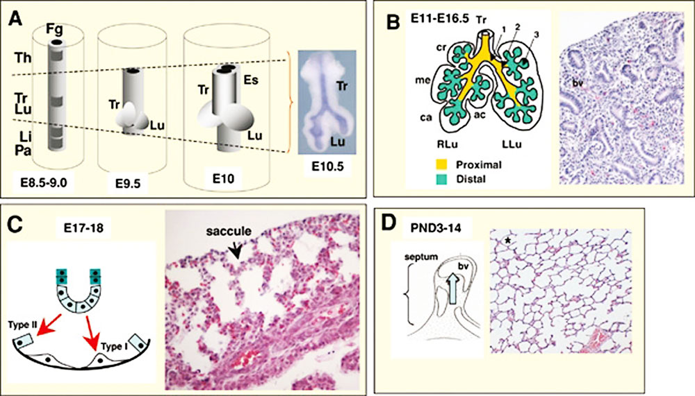
Mouse Respiratory System Development
A. Initial events. From around embryonic day E8.5-9.5 the foregut endoderm (represented in grey) is specified into organ-specific domains, such as the thyroid (Th), trachea (Tr), lung (Lu), liver (Li) and pancreas (Pa). Progenitor cells of the trachea and lung arise from the ventral foregut endoderm in a domain located posterior to the thyroid and anterior to the liver and the pancreatic fields (depicted by dashed lines in A). At E9.5, primary lung buds and the tracheal primordium are induced. From E9.5-E10 primary buds elongate, while the tracheal primordium starts separating from the primitive esophagus (Es), presumably by growth of an ascending tracheoesophageal septum. At E10.5 secondary buds form from the primary lung buds (inset picture in A).
B. From E11-E16.5 the epithelium undergoes branching morphogenesis and proximal-distal patterning to form the bronchial tree (arrows 1, 2, 3, represent primary, secondary and tertiary buds, respectively). This period encompasses the pseudoglandular (B, right panel, Hematoxilin and eosin – H&E- staining of a E15.5 lung section showing a gland-like appearance of the distal region), and the canalicular stage (not shown). The lung is subdivided into morphologic units called lobes, surrounded by mesothelial cells of the pleura. Mice have one lobe on the left lung (LLu) and four lobes on the right lung (RLu); these lobes are the cranial (cr), medial (me), caudal (ca) and accessory (ac) lobes.
C. From around E17-E18 the distal epithelial tubules differentiate into type I and type II cells and expand to form saccules (arrows), the primitive gas exchange region of the lung (E18, H&E, right panel).
D. From around postnatal 3-14 the saccules are subdivided into small units through growth of a secondary septum (septation, or alveolization, diagram on left) to give rise to the mature alveoli (H&E, asterisk in right panel). bv: blood vessel. Modified with permission from Cardoso and Lu (2006).
Original file name: Figure 1. Cardoso01.jpg http://www.ncbi.nlm.nih.gov/bookshelf/br.fcgi?book=stembook&part=specificationandpatterning&rendertype=figure&id=specificationandpatterning.F1
Reference
Cardoso WV, Kotton DN. Specification and patterning of the respiratory system. StemBook [Internet]. Cambridge (MA): Harvard Stem Cell Institute; 2008 Jul 16. PMID20614584 | StemBook - Specification and patterning of the respiratory system
Copyright : © 2008 Wellington V. Cardoso and Darrell N. Kotton.
This is an open-access article distributed under the terms of the Creative Commons Attribution License, which permits unrestricted use, distribution, and reproduction in any medium, provided the original work is properly cited.
File history
Click on a date/time to view the file as it appeared at that time.
| Date/Time | Thumbnail | Dimensions | User | Comment | |
|---|---|---|---|---|---|
| current | 06:41, 11 November 2010 | | 1,000 × 571 (125 KB) | S8600021 (talk | contribs) | ==Mouse Respiratory System Development== A. Initial events. From around embryonic day E8.5-9.5 the foregut endoderm (represented in grey) is specified into organ-specific domains, such as the thyroid (Th), trachea (Tr), lung (Lu), liver (Li) and pancreas |
You cannot overwrite this file.
File usage
The following page uses this file: