File:Model capacitation-induced acrosome docking to sperm membrane.jpg
From Embryology
No higher resolution available.
Model_capacitation-induced_acrosome_docking_to_sperm_membrane.jpg (600 × 489 pixels, file size: 73 KB, MIME type: image/jpeg)
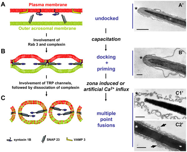
Hypothetical model for capacitation-induced stable docking of the acrosome to the sperm plasma membrane
- A - In control sperm the PM (in red) and OAM (in green) are not associated together. We found syntaxin 1B (purple blue) and VAMP 3 (yellow) at the PM and SNAP 23 (light blue) at the OAM. The undocked PM appears as loose arrangement at the entire head area due to the osmotic effect by EM processing (A′).
- B - In vitro capacitation caused stable docking of the OAM to the apical PM. A stable ternary- SNARE complex is formed but this did not result in exocytotic membrane fusions (B′).
- C - Exocytotic membrane fusions are executed after sperm binding to the zona pellucida or in vitro by calcium ionophore treatments. Mixed vesicles of the apical PM and the OAM are the result of the multipoint fusions characteristic for sperm acrosome exocytosis (C′1). However, at the equatorial sperm head area (distal from the arrows indicated in panel C′2) this fusion does not take place (C′2). This sperm surface area is specifically involved in the later occurring adhesion and fusion processes between the sperm cell and the oocyte leading to fertilization. The asterisks indicate the apical side of the sperm head.
Bar represents 50 nm.
- Links: Spermatozoa Development | Pig Development
Reference
<pubmed>20585455</pubmed>| PLoS One
Copyright
© 2010 Tsai et al. This is an open-access article distributed under the terms of the Creative Commons Attribution License, which permits unrestricted use, distribution, and reproduction in any medium, provided the original author and source are credited.
Original file mane: journal.pone.0011204.g008.png
File history
Click on a date/time to view the file as it appeared at that time.
| Date/Time | Thumbnail | Dimensions | User | Comment | |
|---|---|---|---|---|---|
| current | 23:23, 21 June 2010 | | 600 × 489 (73 KB) | S8600021 (talk | contribs) | Hypothetical model for capacitation-induced stable docking of the acrosome to the sperm plasma membrane. (A): In control sperm the PM (in red) and OAM (in green) are not associated together. We found syntaxin 1B (purple blue) and VAMP 3 (yellow) at the P |
You cannot overwrite this file.
File usage
The following 2 pages use this file: