File:Lymphocyte rosettes EM01-06.jpg

Original file (1,364 × 2,100 pixels, file size: 334 KB, MIME type: image/jpeg)
Human Lymphocytes with Sheep Erythrocyte Rosettes
Human lymphocytes (Thymocytes, T-cells) and Sheep erythrocytes (Red Blood Cells, SRBC).
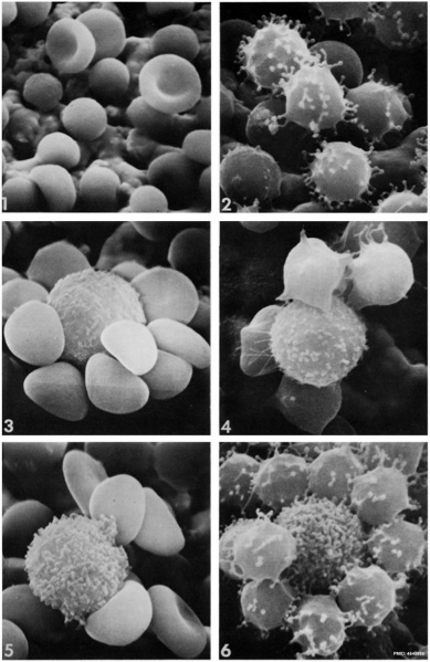
Fig. 1. SRBC washed with HBSS showing spheroechinocyte transformation. Two SRBC have maintained their biconcave shape. X 3,600.
Fig. 2. SRBC showing microspherulation, with multiple microprojections with beaded ends. X 6,900.
Fig. 3. E-rosette showing a smooth surfaced T lymphocyte surrounded by SRBC; the majority of T cells and less than 10% of rosetting B ceils had this type of surface. X 5,700.
Fig. 4. E-rosette showing intermediate type of T lymphocyte.Up to 30% of rosetting B and T cells had this type of surface. X 6,000.
Fig. 5. EAC-rosette, showing villous B cell. The majority of B cells and about 15% of T cells showed this surface morphology.X 5,700.
Fig. 6. Rosetting T lymphocyte with a villous surface, surrounded by spheroechinocyte SRBC which have multiple projections with beaded extremities of similar dimensions to lymphocyte microvilli. This type of picture suggested that deposition of portions of SRBC projections to the lymphocyte surface may occur. X 6,600.
- Lymphocyte EM Images: T and B Lymphocytes 1 TEM | T and B Lymphocytes 2 TEM | T Lymphocyte SEM | B lymphocyte 1 TEM | B lymphocyte 2 TEM | B lymphocyte 3 TEM | Plasma Cell TEM | T2 Lymphocyte 1 TEM | T2 Lymphocyte 2 TEM | lymphocyte rosettes | T lymphocyte 1 | T lymphocyte 2 | T lymphocyte 3 | T lymphocyte 4 | T lymphocyte 5 | T lymphocyte 6 | B lymphocyte | B lymphocytes TEM | Immune System Development | Blood
References
Polliack A, Fu SM, Douglas SD, Bentwich Z, Lampen N & De Harven E. (1974). Scanning electron microscopy of human lymphocyte-sheep erythrocyte rosettes. J. Exp. Med. , 140, 146-58. PMID: 4545895
Copyright
Rockefeller University Press - Copyright Policy This article is distributed under the terms of an Attribution–Noncommercial–Share Alike–No Mirror Sites license for the first six months after the publication date (see http://www.jcb.org/misc/terms.shtml). After six months it is available under a Creative Commons License (Attribution–Noncommercial–Share Alike 4.0 Unported license, as described at https://creativecommons.org/licenses/by-nc-sa/4.0/ ). (More? Help:Copyright Tutorial)
Cite this page: Hill, M.A. (2024, April 20) Embryology Lymphocyte rosettes EM01-06.jpg. Retrieved from https://embryology.med.unsw.edu.au/embryology/index.php/File:Lymphocyte_rosettes_EM01-06.jpg
- © Dr Mark Hill 2024, UNSW Embryology ISBN: 978 0 7334 2609 4 - UNSW CRICOS Provider Code No. 00098G
File history
Click on a date/time to view the file as it appeared at that time.
| Date/Time | Thumbnail | Dimensions | User | Comment | |
|---|---|---|---|---|---|
| current | 12:47, 22 February 2012 | | 1,364 × 2,100 (334 KB) | Z8600021 (talk | contribs) | Sheep Red Blood Cells Fig. 1. SRBC washed with HBSS showing spheroechinocyte transformation. Two SRBC have maintained their biconcaveshape. X 3,600. Fig. 2. SRBC showing microspherulation, with multiple microprojections with beaded ends. X 6,900. Fig. |
You cannot overwrite this file.
File usage
The following 3 pages use this file: