File:Lymph node cartoon 01.jpg
From Embryology
No higher resolution available.
Lymph_node_cartoon_01.jpg (800 × 538 pixels, file size: 109 KB, MIME type: image/jpeg)
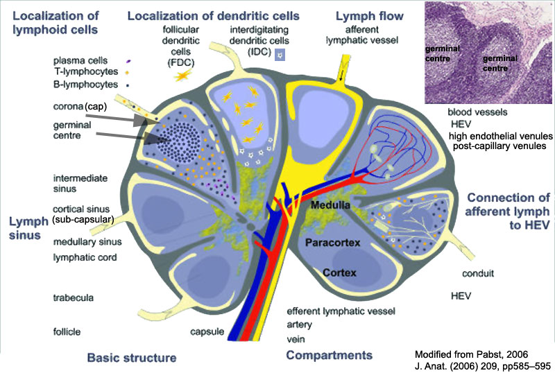
Detailed Lymph Node
- cortex -
- paracortex - a T-cell zone
- medulla -
Lymph pathway
- Afferent vessel
- Subcapsular sinus
- Paratrabecular sinus
- Medullary sinus
- Efferent vessel
Lymphocyte pathway
- high endothelial venule - (HEV) the specialised post-capillary venous region that enables blood lymphocytes to enter a lymph node. The endothelial cells in these venules express ligands that bind lymphocytes, aiding their adhesion and subsequent transmigration into the lymph node.
- Lymph Node Cartoons: Detailed structure | Cartoon with Histology | Lymphocyte traffic | Simple structure | Simple node anatomy | Wiki node image | Internal structure | Mesenteric lymph node | Histology | Gallery | Lymph Node Development
Cite this page: Hill, M.A. (2024, April 18) Embryology Lymph node cartoon 01.jpg. Retrieved from https://embryology.med.unsw.edu.au/embryology/index.php/File:Lymph_node_cartoon_01.jpg
- © Dr Mark Hill 2024, UNSW Embryology ISBN: 978 0 7334 2609 4 - UNSW CRICOS Provider Code No. 00098G
File history
Click on a date/time to view the file as it appeared at that time.
| Date/Time | Thumbnail | Dimensions | User | Comment | |
|---|---|---|---|---|---|
| current | 16:02, 28 February 2011 | | 800 × 538 (109 KB) | S8600021 (talk | contribs) |
You cannot overwrite this file.