File:Lung primary lobule 01.jpg
Original file (800 × 799 pixels, file size: 140 KB, MIME type: image/jpeg)
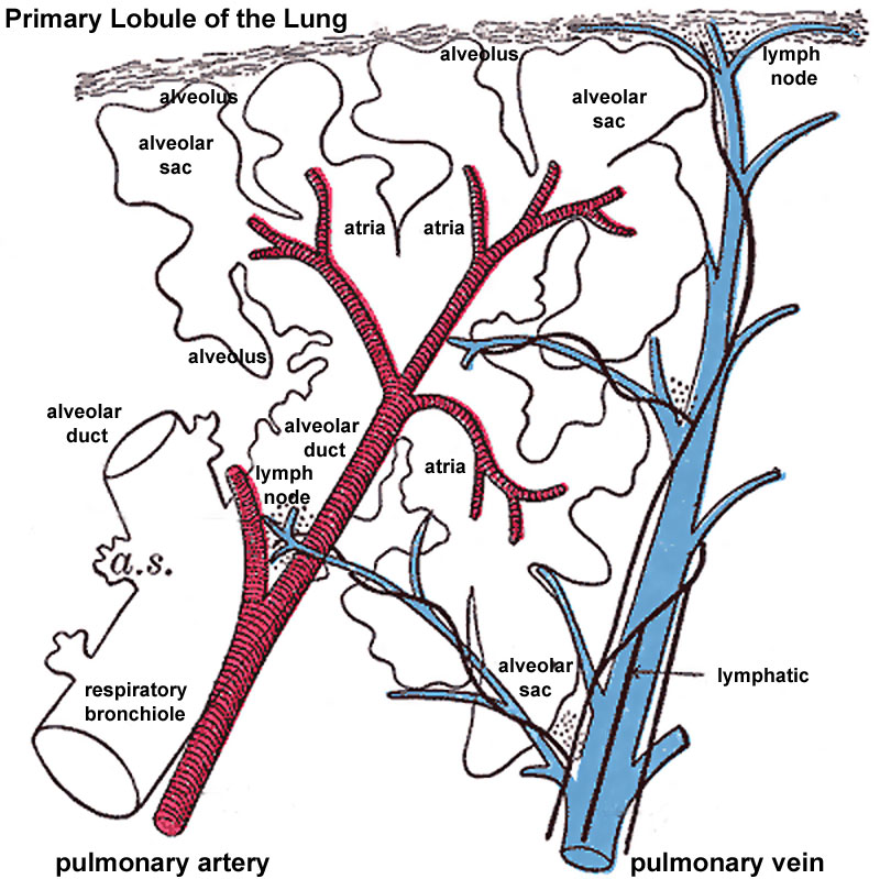
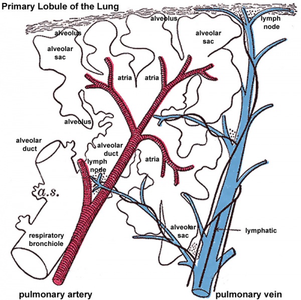
Primary Lung Lobule
Longitudinal Section schematic of the lung anatomical unit (Miller.) Based on Gray's Anatomy Fig.975
respiratory bronchiole ->alveolar duct -> atria -> alveolar sac -> alveolus (plural, alveoli)
- Links: Primary Lung Lobule | Gray 975 | Secondary Lung Lobule | Gray 974 | Respiratory System Development
Alveoli
The alveoli are lined by a delicate layer of simple squamous epithelium, the cells of which are united at their edges by cement substance. Between the squames are here and there smaller, polygonal, nucleated cells. Outside the epithelial lining is a little delicate connective tissue containing numerous elastic fibers and a close net-work of blood capillaries, and forming a common wall to adjacent alveoli.
Vessels and Nerves
The pulmonary artery conveys the venous blood to the lungs; it divides into branches which accompany the bronchial tubes and end in a dense capillary net-work in the walls of the alveoli. In the lung the branches of the pulmonary artery are usually above and in front of a bronchial tube, the vein below.
The pulmonary capillaries form plexuses which lie immediately beneath the lining epithelium, in the walls and septa of the alveoli and of the infundibula. In the septa between the alveoli the capillary net-work forms a single layer. The capillaries form a very minute net-work, the meshes of which are smaller than the vessels themselves; their walls are also exceedingly thin. The arteries of neighboring lobules are independent of each other, but the veins freely anastomose.
The pulmonary veins commence in the pulmonary capillaries, the radicles coalescing into larger branches which run through the substance of the lung, independently of the pulmonary arteries and bronchi. After freely communicating with other branches they form large vessels, which ultimately come into relation with the arteries and bronchial tubes, and accompany them to the hilus of the organ. Finally they open into the left atrium of the heart, conveying oxygenated blood to be distributed to all parts of the body by the aorta.
The bronchial arteries supply blood for the nutrition of the lung; they are derived from the thoracic aorta or from the upper aortic intercostal arteries, and, accompanying the bronchial tubes, are distributed to the bronchial glands and upon the walls of the larger bronchial tubes and pulmonary vessels. Those supplying the bronchial tubes form a capillary plexus in the muscular coat, from which branches are given off to form a second plexus in the mucous coat; this plexus communicates with small venous trunks that empty into the pulmonary veins. Others are distributed in the interlobular areolar tissue, and end partly in the deep, partly in the superficial, bronchial veins. Lastly, some ramify upon the surface of the lung, beneath the pleura, where they form a capillary network.
The bronchial vein is formed at the root of the lung, receiving superficial and deep veins corresponding to branches of the bronchial artery. It does not, however, receive all the blood supplied by the artery, as some of it passes into the pulmonary veins. It ends on the right side in the azygos vein, and on the left side in the highest intercostal or in the accessory hemiazygos vein.
The lymphatics are described elsewhere.
(Text modified from Gray's Anatomy)
File history
Click on a date/time to view the file as it appeared at that time.
| Date/Time | Thumbnail | Dimensions | User | Comment | |
|---|---|---|---|---|---|
| current | 16:59, 29 February 2012 | | 800 × 799 (140 KB) | Z8600021 (talk | contribs) | ==Longitudinal Section of a Primary Lobule of the Lung== Schematic of the lung anatomical unit (Miller.) Based on Gray's Anatomy Fig.975 Category:Respiratory Category:Historic Embryology Category:Cartoon |
You cannot overwrite this file.