File:Limb mesenchymal cell shape.jpg
Original file (775 × 1,000 pixels, file size: 180 KB, MIME type: image/jpeg)
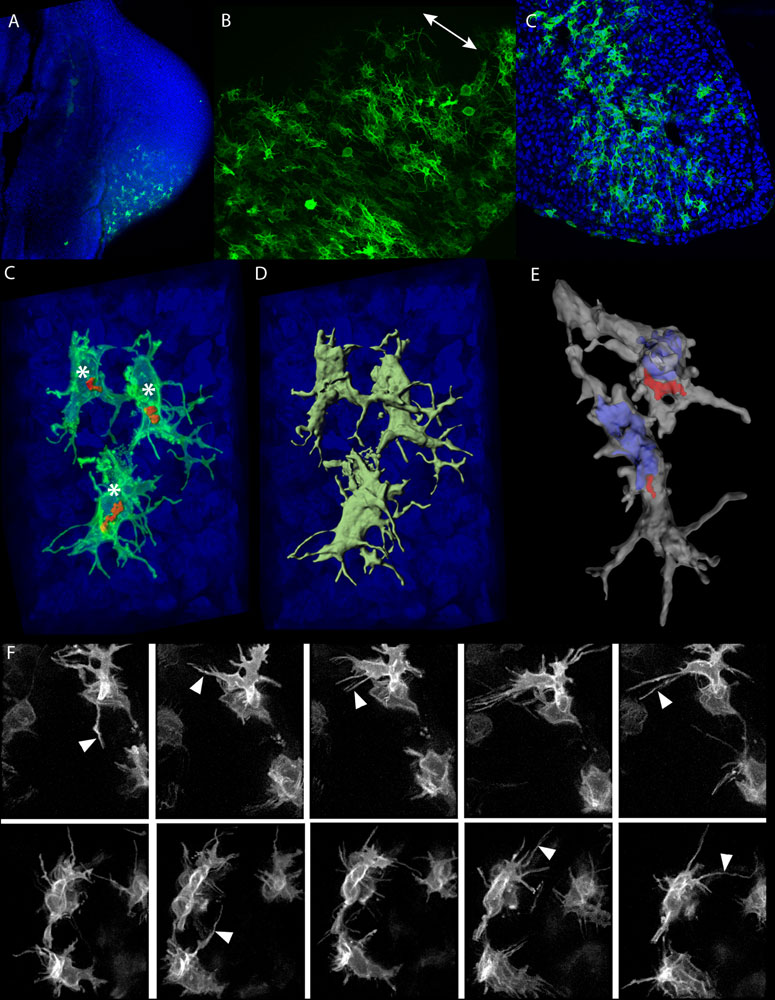
Limb bud mesenchymal cells display complex 3D shapes with highly dynamic filopodia
(A) Overview of a typical in ovo electroporation result. All cell nuclei are labelled with DAPI (blue) and the GFP expression can be seen in green. (B) When many cells are labelled a general alignment of cellular processes is evident, oriented perpendicular (white double arrow) to the nearest ectoderm (bottom-right). (C) A section through the middle of a chick limb bud, showing the complex, branched and extended morphology of almost all the randomly labelling cells. (D–E) High-magnification 3D images of a few labelled cells, showing how the Golgi (red) is consistently on the same side of the nucleus (asterisks in C, blue shape in E) and revealing the extended cellular processes of the complex 3D geometry of each cell. (F) Frames from two time-lapse movies showing the dynamic extension and retraction of the filopodia (white arrowheads) over a 2 h period.
Figure 7. Journal.pbio.1000420.g007.png
doi:10.1371/journal.pbio.1000420.g007
Reference
<pubmed>20644711</pubmed>| PMC2903592 | PLoS
Citation: Boehm B, Westerberg H, Lesnicar-Pucko G, Raja S, Rautschka M, et al. (2010) The Role of Spatially Controlled Cell Proliferation in Limb Bud Morphogenesis. PLoS Biol 8(7): e1000420. doi:10.1371/journal.pbio.1000420
Copyright: © 2010 Boehm et al. This is an open-access article distributed under the terms of the Creative Commons Attribution License, which permits unrestricted use, distribution, and reproduction in any medium, provided the original author and source are credited.
File history
Click on a date/time to view the file as it appeared at that time.
| Date/Time | Thumbnail | Dimensions | User | Comment | |
|---|---|---|---|---|---|
| current | 10:30, 23 March 2011 | | 775 × 1,000 (180 KB) | S8600021 (talk | contribs) | ==Limb bud mesenchymal cells display complex 3D shapes with highly dynamic filopodia== (A) Overview of a typical in ovo electroporation result. All cell nuclei are labelled with DAPI (blue) and the GFP expression can be seen in green. (B) When many cell |
You cannot overwrite this file.
File usage
The following 2 pages use this file: