File:Limb bud geometry and patterning.jpg
Original file (583 × 765 pixels, file size: 76 KB, MIME type: image/jpeg)
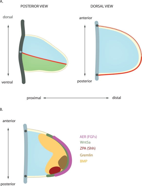
Geometry and Patterning of the Limb Bud
A Geometry of the limb bud.
- Yellow - ectodermal layer.
- Blue - dorsal.
- Green - ventral mesenchyme.
- Red line - indicates the dorsoventral boundary (solid - posterior, dashed - anterior).
- Thick grey lines - represent the flank, with the dots indicating the points of cross-section between the posterior and the dorsal view.
B Expression domains of patterning signals.
Legend
- AER - apical ectodermal ridge, expresses FGF encoding genes (Fgf8, Fgf4, Fgf9, Fgf17).
- ZPA - zone of polarizing activity, is the source of Sonic Hedgehog (Shh). Bone morphogenetic protein (BMP4) is expressed in a broad domain, which is progressively restricted in time. Gremlin1 is a BMP antagonist. BMP, Gremlin, Shh, and FGF are interlinked in signaling feedback loops, which causes their domains of expression and activity to change over time. Wnt5a is expressed in a proximo-distal gradient, with highest levels at the tip of the limb bud.
Original file name: Figure 2. Pbio.1000421.g002.jpg
Reference
<pubmed>20644713</pubmed>| PMC2903596 | PLoS
Citation: Kicheva A, Briscoe J (2010) Limbs Made to Measure. PLoS Biol 8(7): e1000421. doi:10.1371/journal.pbio.1000421
Published: July 13, 2010
Copyright: © 2010 Kicheva, Briscoe. This is an open-access article distributed under the terms of the Creative Commons Attribution License, which permits unrestricted use, distribution, and reproduction in any medium, provided the original author and source are credited.
File history
Click on a date/time to view the file as it appeared at that time.
| Date/Time | Thumbnail | Dimensions | User | Comment | |
|---|---|---|---|---|---|
| current | 11:20, 23 July 2010 | | 583 × 765 (76 KB) | S8600021 (talk | contribs) | ==Geometry and patterning of the limb bud== A) Geometry of the limb bud. Yellow - ectodermal layer. Blue – dorsal. Green - ventral mesenchyme. Red line indicates the dorsoventral boundary (solid - posterior, dashed - anterior). The thick grey lines rep |
You cannot overwrite this file.