File:Lewis1920 fig08.jpg

Original file (1,000 × 765 pixels, file size: 81 KB, MIME type: image/jpeg)
Figure 8
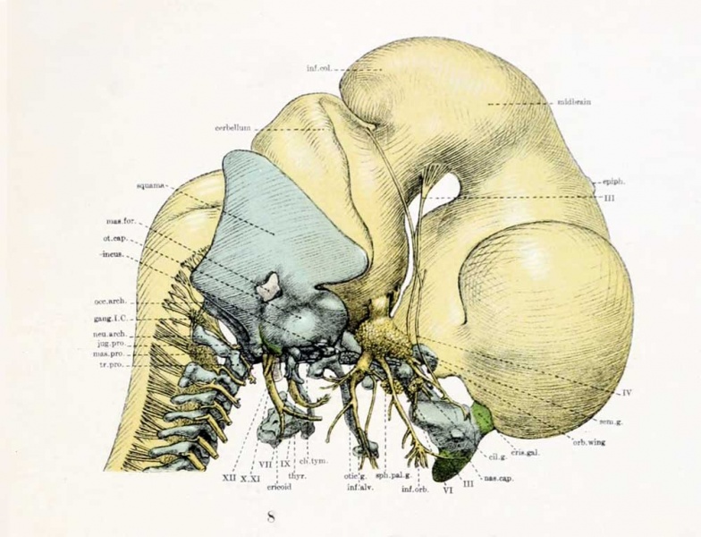
Lateral aspect of cartilaginous skull and cervical vertebra; with the brain, cervical cord, and nerves. X10 diameters.
| Historic Disclaimer - information about historic embryology pages |
|---|
 |
All of the figures except No. 4 are from models or combinations of models and graphic reconstructions of one embryo No. 460, Carnegie Collection. The illustrations are mainly the work of Mr. C. W. Shepard and Mr. J. F. Didusch.
Cartilage is colored blue, except figure 4, precartilage green, nerves yellow, muscles red; the blastema remains uncolored. All magnifications refer to the original published image size.
- Links: Plate 1 | Plate 2 | Plate 3 | Plate 4 | Plate 5 | Abbreviations | 1920 Lewis | Carnegie Embryo 460 | Carnegie stage 20 | Week 8 | Volume IX | Contributions to Embryology | Skull Development
Reference
Lewis WH. The cartilaginous skull of a human embryo twenty-one millimeters in length. (1920) Contrib. Embryol., Carnegie Inst. Wash. Publ. 272, 9: 299-324.
Cite this page: Hill, M.A. (2024, April 25) Embryology Lewis1920 fig08.jpg. Retrieved from https://embryology.med.unsw.edu.au/embryology/index.php/File:Lewis1920_fig08.jpg
- © Dr Mark Hill 2024, UNSW Embryology ISBN: 978 0 7334 2609 4 - UNSW CRICOS Provider Code No. 00098G
File history
Click on a date/time to view the file as it appeared at that time.
| Date/Time | Thumbnail | Dimensions | User | Comment | |
|---|---|---|---|---|---|
| current | 12:12, 14 April 2012 | | 1,000 × 765 (81 KB) | Z8600021 (talk | contribs) | ==Figure 8== Plate 3: Fig. 8 | Fig. 9 {{Lewis1920}} |
You cannot overwrite this file.