File:Kollmann447.jpg
Kollmann447.jpg (564 × 556 pixels, file size: 42 KB, MIME type: image/jpeg)
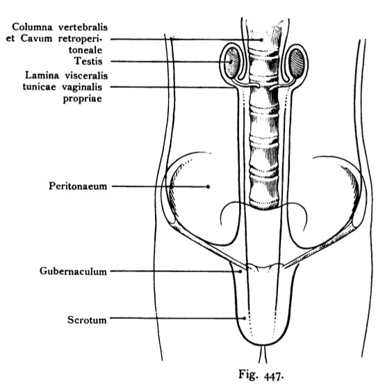
Fig. 447. Descent of testicles
Schema. (After Tillaux.)
The figure represents the abdominal cavity, whose background in the spine
is visible. With blue paint, the peritoneum is indicated. The gonads,
which later constitutes the testis is located in the upper abdomen on both
Sides of the aorta (see figure 429) and the retroperitoneum to the bar
channel descending testicle receives a coating of the peritoneum at its
Enter the abdominal cavity. This coating is called the visceral layer (lamina
visceral) of the tunica vaginalis propria testis (formerly called Tunica adnata GE
called it). Inguinal canal is not yet developed, and no prior vaginal process-
hands.
- This text is a Google translate computer generated translation and may contain many errors.
Images from - Atlas of the Development of Man (Volume 2)
(Handatlas der entwicklungsgeschichte des menschen)
- Kollmann Atlas 2: Gastrointestinal | Respiratory | Urogenital | Cardiovascular | Neural | Integumentary | Smell | Vision | Hearing | Kollmann Atlas 1 | Kollmann Atlas 2 | Julius Kollmann
- Links: Julius Kollman | Atlas Vol.1 | Atlas Vol.2 | Embryology History
| Historic Disclaimer - information about historic embryology pages |
|---|
 |
Reference
Kollmann JKE. Atlas of the Development of Man (Handatlas der entwicklungsgeschichte des menschen). (1907) Vol.1 and Vol. 2. Jena, Gustav Fischer. (1898).
Cite this page: Hill, M.A. (2024, April 19) Embryology Kollmann447.jpg. Retrieved from https://embryology.med.unsw.edu.au/embryology/index.php/File:Kollmann447.jpg
- © Dr Mark Hill 2024, UNSW Embryology ISBN: 978 0 7334 2609 4 - UNSW CRICOS Provider Code No. 00098G
Fig. 447. Descensus testiculorum.
Schema. (Nach Tillaux.)
Die Figur stellt die Bauchhöhle dar, in deren Hintergrund die Wirbelsäule sichtbar ist. Mit blauer Farbe ist das Peritoneum angedeutet. Die Keimdrüse, welche später den Hoden darstellt, liegt oben in der Bauchhöhle zu beiden Seiten der Aorta (vergl. die Figur 429) und retroperitoneaL Der zum Leisten- kanal herabsteigende Hoden erhält einen Überzug des Peritonaeums bei seinem Eintreten in den Bauchraum. Dieser Überzug heißt das viscerale Blatt (Lamina visceralis) der Tunica vaginalis propria testis (früher auch Tunica adnata ge- nannt). Noch ist kein Leistenkanal entwickelt und kein Processus vaginalis vor- handen.
File history
Click on a date/time to view the file as it appeared at that time.
| Date/Time | Thumbnail | Dimensions | User | Comment | |
|---|---|---|---|---|---|
| current | 21:46, 16 October 2011 | | 564 × 556 (42 KB) | S8600021 (talk | contribs) | {{Kollmann1907}} |
You cannot overwrite this file.
File usage
The following 3 pages use this file: