File:Keith1902 fig057.jpg

Original file (887 × 700 pixels, file size: 159 KB, MIME type: image/jpeg)
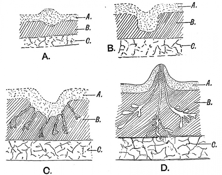
Fig. 57 Showing the various stages in tho development of the Mamma
A = Epiblast. B = Subcutaneous tissue (mesoblast). C = Pectoralis major.
Seven stages may be recognised in the developmental history of the glandular mammary tissue. Four of these take place before birth :
1st (Fig. 57 A). The deeper layer of epiblast thickens over the mammary area ; this thickening represents a part of the mammary ridge or line. This stage is seen in the 2nd month.
2nd (Fig. 57 B). The thickening becomes depressed, thus giving rise to a slight pit on the surface.
3rd (Fig. 57 C). From the depression arises a number of buds, exactly similar to those of sweat gland (5 th month). The stalks of these buds form the epithelial lining of the lactiferous ducts.
4th (Fig. 57 D). The lobular buds, for each bud develops into a lobe, subdivide at their growing extremities. At first solid, they begin to caniculize (7th to 9th months). At or about birth the pit or depression, from which the lobular buds originated, is raised, evaginated and forms the surface of the nipple (Fig. 5 7 D).
| Historic Disclaimer - information about historic embryology pages |
|---|
 |
- The Skin and its Appendages: Fig. 51. Skin strata first month | Fig. 52. Skin strata second month | Fig. 53. Skin strata sixth month onwards | Fig. 54. Common dermal papillae patterns on the finger tips | Fig. 55. Developing Hair | Fig. 56. Diagrammatic Section across a Nail | Fig. 57. Stages in Mamma development | Fig. 58. Section of the Breast | Figures
| Historic Disclaimer - information about historic embryology pages |
|---|
|
Human Embryology and Morphology (1902): Development or the Face | The Nasal Cavities and Olfactory Structures | Development of the Pharynx and Neck | Development of the Organ of Hearing | Development and Morphology of the Teeth | The Skin and its Appendages | The Development of the Ovum of the Foetus from the Ovum of the Mother | The Manner in which a Connection is Established between the Foetus and Uterus | The Uro-genital System | Formation of the Pubo-femoral Region, Pelvic Floor and Fascia | The Spinal Column and Back | The Segmentation of the Body | The Cranium | Development of the Structures concerned in the Sense of Sight | The Brain and Spinal Cord | Development of the Circulatory System | The Respiratory System | The Organs of Digestion | The Body Wall, Ribs, and Sternum | The Limbs | Figures | Embryology History
Reference
Keith A. Human Embryology and Morphology. (1902) London: Edward Arnold.
Cite this page: Hill, M.A. (2024, April 20) Embryology Keith1902 fig057.jpg. Retrieved from https://embryology.med.unsw.edu.au/embryology/index.php/File:Keith1902_fig057.jpg
- © Dr Mark Hill 2024, UNSW Embryology ISBN: 978 0 7334 2609 4 - UNSW CRICOS Provider Code No. 00098G
File history
Click on a date/time to view the file as it appeared at that time.
| Date/Time | Thumbnail | Dimensions | User | Comment | |
|---|---|---|---|---|---|
| current | 09:14, 7 January 2014 | | 887 × 700 (159 KB) | Z8600021 (talk | contribs) |
You cannot overwrite this file.
File usage
The following 3 pages use this file: