File:Keith1902 fig037.jpg
Original file (800 × 668 pixels, file size: 62 KB, MIME type: image/jpeg)
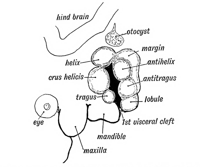
Fig. 37 Showing the Tubercles which arise round the First Visceral Cleft to form the External Ear
Six tubercles appear on the mandibular and hyoid arches round the 1st cleft depression and form the external ear (see Fig. 37 and Fig. 38). Two of these tubercles grow from the mandibular arch and form the tragus and crus of the helix ; three from the hyoid to form the lobule, antitragus from the tissue behind the tubercles which form the antihelix and antitragus.
The auricular tubercles may not fuse completely and thus leave fistulae between them. Such fistulae are commonly seen between the tragus and root of the helix, or between the antihelix and helix. The outgrowth of the ear may be arrested at any stage. The mandibular part is supplied, as one would expect from its origin, by the third division of the 5th, while the sensory fibres of the hyoid part come from the 2nd cervical by the great auricular and small occipital nerves.
- Development of the Organ of Hearing: Fig. 35. Cephalic region of an embryo, showing the origin of the Auditory System | Fig. 36 A. Adult External Auditory Meatus | Fig. 36 B. External Auditory Meatus at Birth | Fig. 37. Tubercles round the First Visceral Cleft to form the External Ear | Fig. 38. Part of the Adult Ear formed by each Tubercle | Fig. 39. Auditory Organs 6th week human fetus | Fig. 40. Cavities from the Inner Recess of the First Cleft | Fig. 41. The temporal bone at birth | Fig. 42. Walls of the Antrum | Fig. 43. Outer aspect of the Petro-mastoid at birth | Fig. 44. Membranous Labyrinth | Fig. 45. The Otocyst in an Embryo of five weeks | Fig. 46. Nerve Structures Sense of Hearing | Figures
| Historic Disclaimer - information about historic embryology pages |
|---|
 |
Human Embryology and Morphology (1902): Development or the Face | The Nasal Cavities and Olfactory Structures | Development of the Pharynx and Neck | Development of the Organ of Hearing | Development and Morphology of the Teeth | The Skin and its Appendages | The Development of the Ovum of the Foetus from the Ovum of the Mother | The Manner in which a Connection is Established between the Foetus and Uterus | The Uro-genital System | Formation of the Pubo-femoral Region, Pelvic Floor and Fascia | The Spinal Column and Back | The Segmentation of the Body | The Cranium | Development of the Structures concerned in the Sense of Sight | The Brain and Spinal Cord | Development of the Circulatory System | The Respiratory System | The Organs of Digestion | The Body Wall, Ribs, and Sternum | The Limbs | Figures | Embryology History
Reference
Keith A. Human Embryology and Morphology. (1902) London: Edward Arnold.
Cite this page: Hill, M.A. (2024, April 18) Embryology Keith1902 fig037.jpg. Retrieved from https://embryology.med.unsw.edu.au/embryology/index.php/File:Keith1902_fig037.jpg
- © Dr Mark Hill 2024, UNSW Embryology ISBN: 978 0 7334 2609 4 - UNSW CRICOS Provider Code No. 00098G
File history
Click on a date/time to view the file as it appeared at that time.
| Date/Time | Thumbnail | Dimensions | User | Comment | |
|---|---|---|---|---|---|
| current | 20:02, 29 December 2013 | | 800 × 668 (62 KB) | Z8600021 (talk | contribs) | {{Keith1902_4_figures}} {{Human embryology morphology 1902 footer}} |
You cannot overwrite this file.
File usage
The following 3 pages use this file: