File:Keith1902 fig036b.jpg

Original file (678 × 639 pixels, file size: 70 KB, MIME type: image/jpeg)
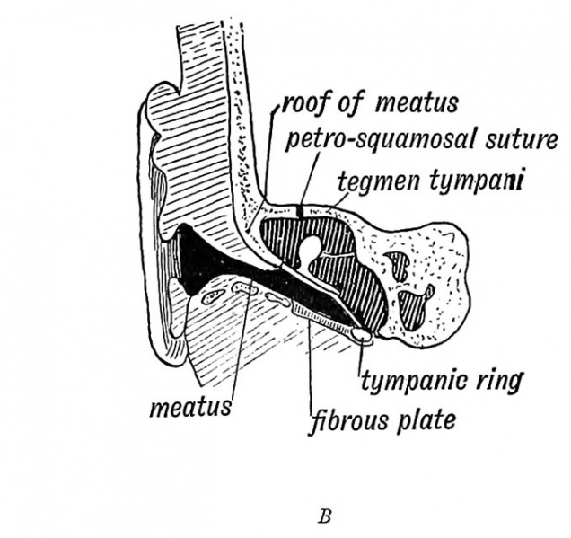
Fig. 36 B. Section of the External Auditory Meatus at Birth
(After Symington.)
The external auditory meatus is derived from the upper part of the first cleft depression. In the adult the meatus is 1.25 in. long ; at birth it is 0.33 of an inch, and is surrounded by fibro-cartilaginous and fibrous tissue. In the adult the tympanic ring grows outwards in the fibrous tissue, as we have already seen (page IV) to form the tympanic plate and the inner two thirds of the meatal floor. The squamous part of the temporal, which is developed over in its roof, also grows outwards and forms a thick, horizontal plate in the inner two-thirds of the meatal roof (Figs. 36 A and 36 B). Over the roof lies the third temporal convolution.
The meatus is supplied in front by the nerve of the mandibular arch (auriculo-temporal branch). Why the vagus should supply it with a branch (Arnold's nerve) is obscure. The vagus is a visceral nerve and supplies the 3rd and 4th clefts by its superior and inferior laryngeal branches. In fishes a branch of the vagus passes backwards beneath the skin on each side and supplies the sense organs of the lateral line. Many regard the auricular branch of the vagus as a vestige of such a branch.
In the newly-born child the membrana tympani is so obliquely set that its outer surface is almost in contact with the meatal floor. With the development in length of the meatus, it becomes more vertical in position. The meatus may be only partly developed or even absent, the upper part of the 1st cleft becoming completely closed like the lower part. In such a case there is commonly a corresponding absence of development of the middle and internal ear.
- Development of the Organ of Hearing: Fig. 35. Cephalic region of an embryo, showing the origin of the Auditory System | Fig. 36 A. Adult External Auditory Meatus | Fig. 36 B. External Auditory Meatus at Birth | Fig. 37. Tubercles round the First Visceral Cleft to form the External Ear | Fig. 38. Part of the Adult Ear formed by each Tubercle | Fig. 39. Auditory Organs 6th week human fetus | Fig. 40. Cavities from the Inner Recess of the First Cleft | Fig. 41. The temporal bone at birth | Fig. 42. Walls of the Antrum | Fig. 43. Outer aspect of the Petro-mastoid at birth | Fig. 44. Membranous Labyrinth | Fig. 45. The Otocyst in an Embryo of five weeks | Fig. 46. Nerve Structures Sense of Hearing | Figures
| Historic Disclaimer - information about historic embryology pages |
|---|
 |
Human Embryology and Morphology (1902): Development or the Face | The Nasal Cavities and Olfactory Structures | Development of the Pharynx and Neck | Development of the Organ of Hearing | Development and Morphology of the Teeth | The Skin and its Appendages | The Development of the Ovum of the Foetus from the Ovum of the Mother | The Manner in which a Connection is Established between the Foetus and Uterus | The Uro-genital System | Formation of the Pubo-femoral Region, Pelvic Floor and Fascia | The Spinal Column and Back | The Segmentation of the Body | The Cranium | Development of the Structures concerned in the Sense of Sight | The Brain and Spinal Cord | Development of the Circulatory System | The Respiratory System | The Organs of Digestion | The Body Wall, Ribs, and Sternum | The Limbs | Figures | Embryology History
Reference
Keith A. Human Embryology and Morphology. (1902) London: Edward Arnold.
Cite this page: Hill, M.A. (2024, April 20) Embryology Keith1902 fig036b.jpg. Retrieved from https://embryology.med.unsw.edu.au/embryology/index.php/File:Keith1902_fig036b.jpg
- © Dr Mark Hill 2024, UNSW Embryology ISBN: 978 0 7334 2609 4 - UNSW CRICOS Provider Code No. 00098G
File history
Click on a date/time to view the file as it appeared at that time.
| Date/Time | Thumbnail | Dimensions | User | Comment | |
|---|---|---|---|---|---|
| current | 19:59, 29 December 2013 | | 678 × 639 (70 KB) | Z8600021 (talk | contribs) | {{Keith1902_4_figures}} {{Human embryology morphology 1902 footer}} |
You cannot overwrite this file.
File usage
The following 4 pages use this file: