File:Keibel Mall 309.jpg
Keibel_Mall_309.jpg (687 × 479 pixels, file size: 52 KB, MIME type: image/jpeg)
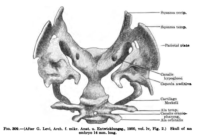
Fig. 309 Human Embryo Skull
Membranous skull of an embryo 14 mm long.
The tissue of the capsules of the labyrinth increases in amount as the labyrinth becomes differentiated. The tissue which encloses the region of the semicircular canals and the vestibule forms an oval mass the outlines of which do not conform to that of the enclosed canals (Fig. 309). This tissue is less dense than most parts of the membranous skeleton of the head and at an early period becomes transformed into embryonic cartilage (see p. 407). The cochlear portion of the labyrinth (Fig. 310) is enclosed by a dense mesenchyme which becomes converted into cartilage at a later period. lateral from the nasal fossa the tissue becomes generally somewhat condensed, though less so than the tissue in the septum. In the perinasal tissue condensation gradually marks out the lateral and ventral portions of the nasal capsule and the membranous floor of the ethmoidal and orbital portions of the cranial cavity (Fig. 310). From the lateral wall membranous processes project into the nasal fossa. These are the anlages of the conchae.
- KM Figure Links: The Germ Cells | Segmentation | First Primitive Segment | Gastrulation | External Form | Placenta | Axial Skeleton | Limb Skeleton | Skull | Muscular System
| Historic Disclaimer - information about historic embryology pages |
|---|
 |
Glossary Links
- Glossary: A | B | C | D | E | F | G | H | I | J | K | L | M | N | O | P | Q | R | S | T | U | V | W | X | Y | Z | Numbers | Symbols | Term Link
Cite this page: Hill, M.A. (2024, April 16) Embryology Keibel Mall 309.jpg. Retrieved from https://embryology.med.unsw.edu.au/embryology/index.php/File:Keibel_Mall_309.jpg
- © Dr Mark Hill 2024, UNSW Embryology ISBN: 978 0 7334 2609 4 - UNSW CRICOS Provider Code No. 00098G
File history
Click on a date/time to view the file as it appeared at that time.
| Date/Time | Thumbnail | Dimensions | User | Comment | |
|---|---|---|---|---|---|
| current | 09:00, 27 August 2012 | | 687 × 479 (52 KB) | Z8600021 (talk | contribs) | ==Fig. 309 Human Embryo Skull== Membranous skull of an embryo 14 mm long. {{Keibel_Mall Images}} Category:Human Category:Human Embryo Category:Musculoskeletal Category:Bone Category:Skull |
You cannot overwrite this file.
File usage
The following 4 pages use this file: