File:Keibel Mall 2 658b.jpg
Original file (895 × 1,200 pixels, file size: 98 KB, MIME type: image/jpeg)
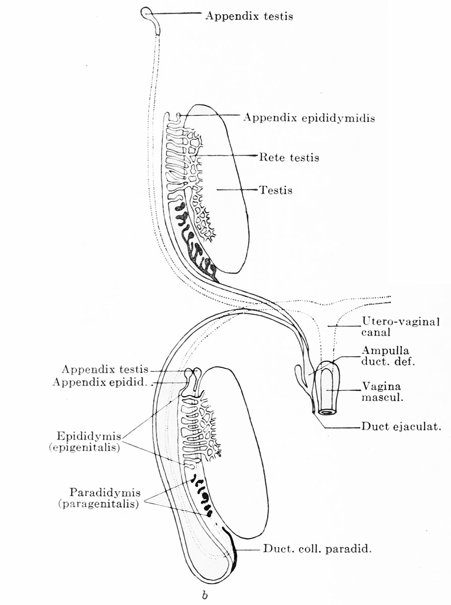
Fig. 658b. Male Genital - After the migration in the male embryo
The testis wanders out of the body cavity into the scrotal sack (descensus). The Mullerian duct degenerates throughout the greater part of its extent, the closed ostium abdominale persisting as the appendix testis (hydatid of the testis) , and the lowest portion of the utero-vaginal canal as the vagina masculina. All the tubules of the epididymis are not employed in the urogenital union: the first tubule remains free and becomes the appendix epididymis (hydatid of the epididymis) and the other unemployed tubules persist as appendices retis. The paradidymis (paragenitalis) is separated into its individual parts, some of the ductuli collectivi persist as the organ of Giraldes and the terminal portions of the ductuli, united to form a canal, persist as the ductus collectivus paradidymidis (ductus aberrans Halleri).
- XIX. - V. Development of External Genitalia: Fig 658a. Male and Female renal and reproductive | Fig 658b. Male Genital | Fig 658b. Female Genital | XIX. The Development of the Urinogenital Organs | Testis Development | Ovary Development | Uterus Development
| Embryology - 24 Apr 2024 |
|---|
| Google Translate - select your language from the list shown below (this will open a new external page) |
|
العربية | català | 中文 | 中國傳統的 | français | Deutsche | עִברִית | हिंदी | bahasa Indonesia | italiano | 日本語 | 한국어 | မြန်မာ | Pilipino | Polskie | português | ਪੰਜਾਬੀ ਦੇ | Română | русский | Español | Swahili | Svensk | ไทย | Türkçe | اردو | ייִדיש | Tiếng Việt These external translations are automated and may not be accurate. (More? About Translations) |
Felix W. The development of the urinogenital organs. In Keibel F. and Mall FP. Manual of Human Embryology II. (1912) J. B. Lippincott Company, Philadelphia. pp 752-979.
| Historic Disclaimer - information about historic embryology pages |
|---|
 |
Cite this page: Hill, M.A. (2024, April 24) Embryology Keibel Mall 2 658b.jpg. Retrieved from https://embryology.med.unsw.edu.au/embryology/index.php/File:Keibel_Mall_2_658b.jpg
- © Dr Mark Hill 2024, UNSW Embryology ISBN: 978 0 7334 2609 4 - UNSW CRICOS Provider Code No. 00098G
Manual of Human Embryology II: Nervous System | Chromaffin Organs and Suprarenal Bodies | Sense-Organs | Digestive Tract and Respiration | Vascular System | Urinogenital Organs | Figures 2 | Manual of Human Embryology 1 | Figures 1 | Manual of Human Embryology 2 | Figures 2 | Franz Keibel | Franklin Mall | Embryology History
Cite this page: Hill, M.A. (2024, April 24) Embryology Keibel Mall 2 658b.jpg. Retrieved from https://embryology.med.unsw.edu.au/embryology/index.php/File:Keibel_Mall_2_658b.jpg
- © Dr Mark Hill 2024, UNSW Embryology ISBN: 978 0 7334 2609 4 - UNSW CRICOS Provider Code No. 00098G
File history
Click on a date/time to view the file as it appeared at that time.
| Date/Time | Thumbnail | Dimensions | User | Comment | |
|---|---|---|---|---|---|
| current | 21:34, 9 February 2014 | | 895 × 1,200 (98 KB) | Z8600021 (talk | contribs) | ==Fig. 658b. == {{Human Embryology Manual 2 19}} {{Keibel_Mall 2 Images}} |
You cannot overwrite this file.
File usage
The following 4 pages use this file: