File:Keibel Mall 2 044.jpg
Original file (889 × 800 pixels, file size: 121 KB, MIME type: image/jpeg)
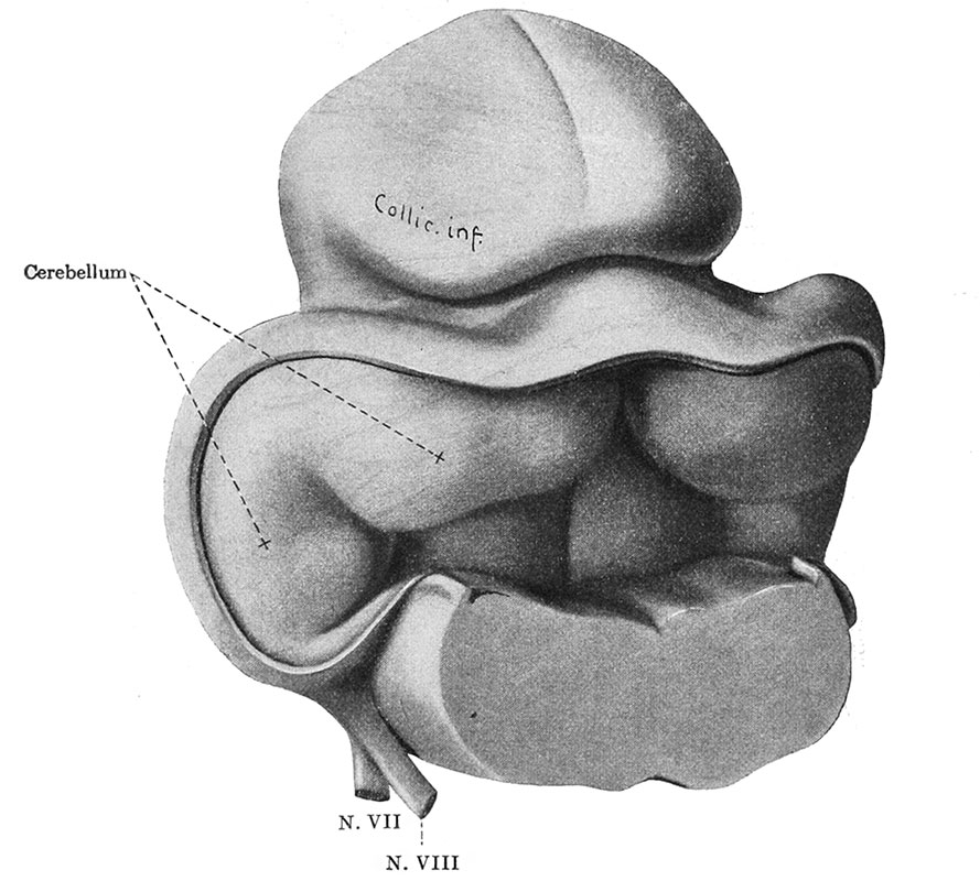
Fig. 44. Reconstruction showing cephalic portion of the rhombencephalon and adjoining midbrain at end of second month
Human embryo 30 mm. long (Mall collection. No. 86). The thickened alar plates form the anlage of the cerebellum and the two halves are still separate. Caudally they are continuous with the nucleus of the acoustic nerve.
| Embryology - 19 Apr 2024 |
|---|
| Google Translate - select your language from the list shown below (this will open a new external page) |
|
العربية | català | 中文 | 中國傳統的 | français | Deutsche | עִברִית | हिंदी | bahasa Indonesia | italiano | 日本語 | 한국어 | မြန်မာ | Pilipino | Polskie | português | ਪੰਜਾਬੀ ਦੇ | Română | русский | Español | Swahili | Svensk | ไทย | Türkçe | اردو | ייִדיש | Tiếng Việt These external translations are automated and may not be accurate. (More? About Translations) |
Streeter GL. The Development of the Nervous System. (1912) chapter 14, vol. 2, in Keibel F. and Mall FP. Manual of Human Embryology II. (1912) J. B. Lippincott Company, Philadelphia.
| Historic Disclaimer - information about historic embryology pages |
|---|
 |
Manual of Human Embryology II: Nervous System | Chromaffin Organs and Suprarenal Bodies | Sense-Organs | Digestive Tract and Respiration | Vascular System | Urinogenital Organs | Figures 2 | Manual of Human Embryology 1 | Figures 1 | Manual of Human Embryology 2 | Figures 2 | Franz Keibel | Franklin Mall | Embryology History
Cite this page: Hill, M.A. (2024, April 19) Embryology Keibel Mall 2 044.jpg. Retrieved from https://embryology.med.unsw.edu.au/embryology/index.php/File:Keibel_Mall_2_044.jpg
- © Dr Mark Hill 2024, UNSW Embryology ISBN: 978 0 7334 2609 4 - UNSW CRICOS Provider Code No. 00098G
File history
Click on a date/time to view the file as it appeared at that time.
| Date/Time | Thumbnail | Dimensions | User | Comment | |
|---|---|---|---|---|---|
| current | 12:08, 24 January 2014 | | 889 × 800 (121 KB) | Z8600021 (talk | contribs) | {{Human Embryology Manual 2 14}} {{Keibel_Mall 2 Images}} |
You cannot overwrite this file.
File usage
The following 2 pages use this file: