File:Keibel Mall 2 030.jpg

Original file (804 × 1,000 pixels, file size: 99 KB, MIME type: image/jpeg)
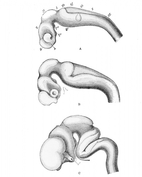
Fig. 30. Profile views of the brains of human embryos at third, fourth, and eighth weeks
Profile as seen during the third (A), fourth (B), and eighth (C) weeks, showing the conversion of the three primary cerebral vesicles into their chief subdivisions and the formation of the flexures of the neural tube. (After Wilhelm His (1831-1904))
A, optic vesicle; Br, pontine region; Gb, auditory vesicle; H, telencephalon; Hb, metencephalon; J, isthmus; M, mesencephalon; N, myelencephalon; NK, neck bend; Pm, mammillary recess; Rf, posterior medullary velum; Tr, infundibular recess; Z, diencephalon.
The formation of the flexures of the neural tube is shown in Fig. 30. It will be seen that there are three distinct flexures, cephalic, pontine and cervical. Two of them have already been mentioned. The third, or cervical flexure, marks the junction of brain and spinal cord and is formed about the same time as the pontine flexure. They are formed, in part at least, in consequence of unequal growth of different parts of the neural tube. They probably influence and also are influenced by the growth of the surrounding structures. The cephalic and cervical flexures involve the surrounding structures to a considerable extent so that there is a corresponding bend of the axis of the whole head, and thus the presence of them can be recognized on the exterior of the embryo. The pontine flexure, however, is limited to the nervous system. The cephalic flexure persists into adult life. The pontine flexure finally disappears and the cervical flexure nearly does.
| Historic Disclaimer - information about historic embryology pages |
|---|
 |
CNS Figures: 22 - neural-plate stage | 23 - 5 to 14 somites stage | 24 - 3.2 mm embryo | 30 - brain 3, 4 and 8 weeks | 32 - 10 mm cranial nerves
- XIV. The Development of the Nervous System: The Histogenesis of Nervous Tissue | The Development of the Central Nervous System | The Peripheral Nervous System | The Sympathetic Nervous System | Manual of Human Embryology II
Cite this page: Hill, M.A. (2024, April 20) Embryology Keibel Mall 2 030.jpg. Retrieved from https://embryology.med.unsw.edu.au/embryology/index.php/File:Keibel_Mall_2_030.jpg
- © Dr Mark Hill 2024, UNSW Embryology ISBN: 978 0 7334 2609 4 - UNSW CRICOS Provider Code No. 00098G
File history
Click on a date/time to view the file as it appeared at that time.
| Date/Time | Thumbnail | Dimensions | User | Comment | |
|---|---|---|---|---|---|
| current | 10:45, 24 January 2014 | | 804 × 1,000 (99 KB) | Z8600021 (talk | contribs) | {{Keibel_Mall 2 Images}} |
You cannot overwrite this file.
File usage
The following 4 pages use this file: