File:Keibel1900 plate01.jpg
Original file (1,952 × 2,451 pixels, file size: 519 KB, MIME type: image/jpeg)
Plate 1
Figure legends text shown below is a slightly modified poor online translation. Original German text.
| Translation Request |
|---|
 If you would like to help with the online translation into English please contact me. |
| Historic Disclaimer - information about historic embryology pages |
|---|
|
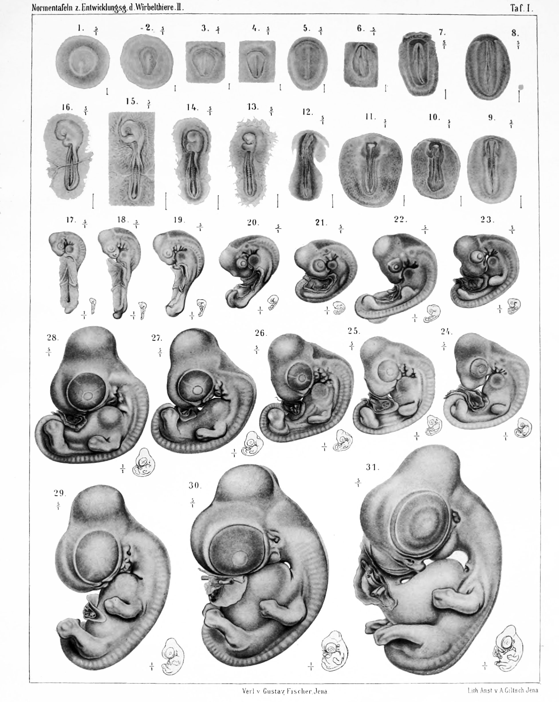
Figure 1
(S.N. 452) The germ shown as Figure 1 is taken from a 9 hours incubated egg fixed with sublimate-acetic acid. The whole seed is approximately circular, its diameter is 4.1 mm. The same in the middle is the zona pellucida, which is near the circular shape, but narrows somewhat towards the rear. Its length is 2.3 mm, and its maximum width 2.15 mm. The primitive streak begins about the middle of the zona pellucida with a button-shaped widening and gradually disappears towards the rear end of the zona pellucida, here a considerable width reaching. Its length is 1.2 mm.
The series shows an early stage of mesoderm formation, the mesoderm does not exceed even the scope of the area pellucida. The primitive streak is already embodied in the typical district front than in the rear. A clear head extension does not exist, nor any traces of blood formation.
Figure 2
(S.N. 436) Also the seed shown in Figure 2 was taken from a 9 hours incubated egg fixed with sublimate-acetic acid. The diameter of the whole seed is 4.2 mm. The shape of the area pellucida is pear-shaped, its length is 2.1 mm, its greatest width 1.5 mm. The primitive streak is 1.6 mm long. Before the front end of primitive streak are cells that create the endoderm is tight and the cover the investments of the head extension well. On the primitive streak a distance is far to detect a shallow trough. At the rear end of the primitive streak area is very wide. The mesoderm not go beyond the area of the area pellucida. No traces of blood formation.
Figure 3
(S.N. 455) Of the seed shown in Figure 3 was taken from a 12 hours incubated egg. The blastoderm had a diameter of about 5 mm. The area pellucida was pear-shaped, but very stocky shape. Its length is 2.3 mm , its greatest width 1.85 mm . The length of the primitive streak is about 1.5 mm. The anterior part of the primitive streak is barely visible in the surface image, in contrast, there are very good as it widens towards the rear . The sagittal shows a front verlötheten with the endoderm head extension , which merges without specific delimitation in the lateral mesoderm. At the front end of the area pellucida I see an irregular band in the surface pattern, which thereby comes to appearance that in its domain, the endoderm is considerably thickened . It is Duval 's " croissant anterieur " , the front sickle The mesoderm is not yet even in this seed over the area of the area pellucid addition. No traces of blood formation.
Figure 4
(S.N. 444) The area shown in Figure 4 pellucida was removed from a 12 hour incubated egg and become fixed in sublimate-acetic acid. It shows a very regular shape, its length is 2.1 mm, its greatest width 1.4 mm. The length of the primitive streak is 1.6 mm. The primitive streak carries a rearwardly significant depth primitive groove that suddenly stops at the rear end. Before the area pellucida a front crescent (croissant anterieur) is available. Microscopic examination shows that the head appendage down with the endoderm in communication and laterally passes into mesodermal cell masses, which also in turn can not be bound, against the endoderm. The mesoderm begins the territory of the area pellucida to pass. From hematopoiesis nothing can be seen.
Figure 5 and 5a
(S.N. 336)
Figures 5 and 5 A are drawn by a blastoderm , which is taken from a 24 hour incubated egg. The boundaries of the area pellucida were not clearly seen in the surface image by reflected light , on the other hand came the peripheral boundaries of the mesoderm , in which the first plants of blood islands are present here , to advantage. The length of the area pellucida is 3 mm , its greatest width 1.9 mm. The length of the primitive streak is 2.8 mm. So the primitive streak reaches almost the tip of the zona pellucida . I have the picture Figure 5 here , where much of the supplementary figure (5a ) for which is drawn according to the same blastoderm at lo - fold magnification of the lower side . The primitive streak and especially the first plant of the vascular Court occur very clearly here . Microscopic examination shows that the mesoderm has spread over the area of the area pellucida . In the area of macroscopically as clearly protruding vessel court to find the first installations of the blood islands . The fact that these arise from the mesoderm , you can , if you compare this stage and slightly older, in which the actual embryonic plant and the first somites have occurred, little doubt .
In summary, I notice too the given of the primitive streak stages pictures that the variation how his time has already highlighted v. Baer, just at this stage is very eye-catching . Baer says (1st c . ) P . 147 and 148 that, the younger embryos would compare " to the more differences and in proportion to the small training to the more important translucent we would become aware of it. This is very striking for the first formation , and all observers to make this remark . Would embryos of the level of education , where the back (ie, the neural canal ) closes [ as well, but ] increases up to the Maasse of adults, drawn on a blackboard next to each other , one would , quite apart from the more rapid or slower progression of the whole development, identify the main differences and believe that these embryos could not develop to the same form itself . Soon, the ratio of head to trunk in an individual is much greater than in the other , and soon the embryos with the exception of the vertebral side of the system of vortices are as transparent as glass , now they are much darker. Some are more curved or more raised from the Keimnaut than others. In some you will not see dre eddy page up to the end of the body, in other , the abdominal plates will already be in the whole extent indicated . Even greater are the differences , if we go back further , and I've been in the narrative of the history of development of the chick ( § ii ) alerted designed how different the primitive streak . Since the formation is still set to such a low level of development that one sees nothing more than surveys and beads so that very reason appear the greater the differences , and one can hardly conceive how these differences lead to the same results and how not next to perfect chickens countless cripples arise . However, since the number of cripples among the older embryos and adult chickens is very low , so you have to back conclude that the differences are compensated , and any deviation is so much möghch routed back to normal. "
Figure 6 and 6a
(S.N. 453; Tab. 3.)
The blastoderm, are drawn after the 6 and 6 a, was taken from a 24 hour incubated egg fixed in sublimate-acetic acid. The germ was characterizes by the first occurrence of medullary bulges, these encompass the caudal very long, slightly curved primitive streak. The investment of the vessel court and the blood islands is also seen. The cranial end of the embryo begins to just stand out from the blastoderm.
Figure 7 and 7a
(S.N. 338; Tab. 4.) The embryo shown in Figures 7 and 7a is taken from a 24 hour incubated egg and fixed in sublimate-acetic acid. The surface picture you can see two somites, the series shows that already 3 are available. Both the systems of medullary folds as the primitive streak have grown considerably in the length. Before the head end takes the mesoderm free point, the facility of proamnions, clearly.
Figure 8 and 8a
(S.N. 357; Tab. 9.) The 20 hours incubated embryo is remarkably well developed, so that one can remember that the egg may have been removed immediately after laying under the hen. The investigation showed that the embryo but still was quite normal. The primitive streak is still very long, but the medullary system begins to gain the upper hand. The medullary folds lie some distance fixed at each other, but still show no organic context. Even with surface observation to 4 somites reveal. The convergence of medullary folds at each place in two places instead, namely in the somites area, but there not yet touching the medullary folds, and then further cranially, where a fixed juxtaposition has already occurred.
Figure 9, 9a and 9b
(S.N. 340; Tab. 16.)
The embryo shown in Figures 9, 9a and 9b is a 24 hour incubated egg. Is was also has advanced for the time relatively far in development. The closure of the medullary folds has now become a partly definite, at other points the medullary folds are separated in this embryo each other only. The primitive streak is still quite long. The surface image of 7 somites can be seen, an 8th is how the series shows. The cranial part of the medullary folds you can see the facilities of the optic vesicles. The anterior intestinal-bay is quite deep. The heart endothelial tubes are still consistently paired. The investment of the vessel court extends a little beyond the cranial end of the embryo.
| Historic Disclaimer - information about historic embryology pages |
|---|
|
Reference
Franz Keibel, Normentafeln zur Entwicklungsgeschichte der Wirbelthiere (Normal plates of the development of vertebrates) Volume Hft.2 (1900) Jena, G. Fischer, Germany.
Cite this page: Hill, M.A. (2024, April 25) Embryology Keibel1900 plate01.jpg. Retrieved from https://embryology.med.unsw.edu.au/embryology/index.php/File:Keibel1900_plate01.jpg
- © Dr Mark Hill 2024, UNSW Embryology ISBN: 978 0 7334 2609 4 - UNSW CRICOS Provider Code No. 00098G
File history
Click on a date/time to view the file as it appeared at that time.
| Date/Time | Thumbnail | Dimensions | User | Comment | |
|---|---|---|---|---|---|
| current | 23:26, 20 November 2013 | | 1,952 × 2,451 (519 KB) | Z8600021 (talk | contribs) |
You cannot overwrite this file.
File usage
The following 3 pages use this file: