File:Jejunum histology 01.jpg
From Embryology
No higher resolution available.
Jejunum_histology_01.jpg (480 × 600 pixels, file size: 79 KB, MIME type: image/jpeg)
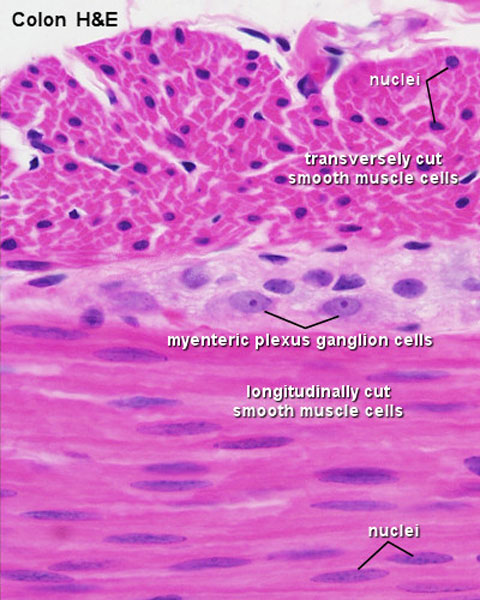
Jejunum Histology
- outer part of the tube of the intestines consists of two layers of smooth muscle.
- one circular layer.
- one longitudinal layer.
- look at the tissue close to the border between the two layers of smooth muscle, you will be able to see both longitudinally sectioned smooth muscle cells and transversely sectioned smooth muscle cells.
- smooth muscle cells are much longer than their nuclei.
- transversely sectioned smooth muscle cells may not have their nuclei in the plane of the section.
- the myenteric plexus can be seen lying between the two smooth muscle layers.
- baboon - H&E
Links: Histology | Histology Stains | Blue Histology images copyright Lutz Slomianka 1998-2009. The literary and artistic works on the original Blue Histology website may be reproduced, adapted, published and distributed for non-commercial purposes. See also the page Histology Stains.
Cite this page: Hill, M.A. (2024, April 20) Embryology Jejunum histology 01.jpg. Retrieved from https://embryology.med.unsw.edu.au/embryology/index.php/File:Jejunum_histology_01.jpg
- © Dr Mark Hill 2024, UNSW Embryology ISBN: 978 0 7334 2609 4 - UNSW CRICOS Provider Code No. 00098G
Original File Name: Jej004he.jpg
File history
Click on a date/time to view the file as it appeared at that time.
| Date/Time | Thumbnail | Dimensions | User | Comment | |
|---|---|---|---|---|---|
| current | 14:15, 6 March 2012 | | 480 × 600 (79 KB) | Z8600021 (talk | contribs) | ==Jejunum Histology-- * baboon - H&E * outer part of the tube forming the intestines consists of two layers of smooth muscle ** one circular layer ** one longitudinal layer. * look at the tissue close to the border between the two layers of smooth muscl |
You cannot overwrite this file.
File usage
The following 2 pages use this file: