File:Intestine histology 001.jpg
From Embryology
No higher resolution available.
Intestine_histology_001.jpg (450 × 600 pixels, file size: 65 KB, MIME type: image/jpeg)
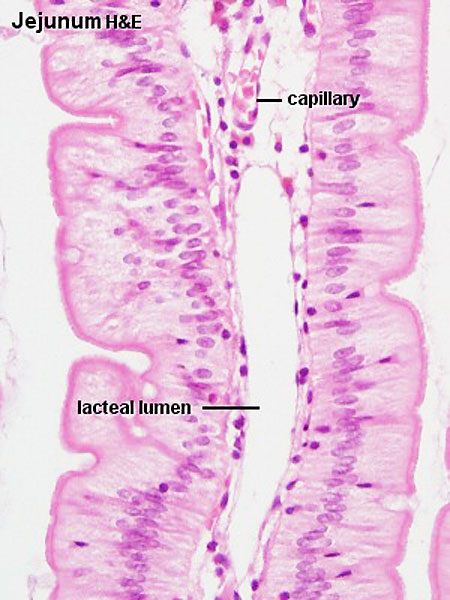
Jejunum
- Baboon, jejunum (Stain - Haematoxylin Eosin)
- cross-section of villi extending into the lumen.
- lacteal - lymphatic capillary surrounded by a layer of flattened endothelial, no blood cells in lumen.
- chyle - milky fluid consisting of lymph and emulsified fat extracted from chyme by the lacteals during digestion and passed to the bloodstream through the thoracic duct.
- chyme - semifluid, creamy material produced by digestion of food.
- Intestine Histology Links: Duodenum overview | Duodenum villi and crypts | Duodenum | Jejunum overview | Jejunum villus | Jejunum labeled | Jejunum unlabeled | Gastrointestinal Tract Histology | Intestine Development
Links: Histology | Histology Stains | Blue Histology images copyright Lutz Slomianka 1998-2009. The literary and artistic works on the original Blue Histology website may be reproduced, adapted, published and distributed for non-commercial purposes. See also the page Histology Stains.
Cite this page: Hill, M.A. (2024, April 25) Embryology Intestine histology 001.jpg. Retrieved from https://embryology.med.unsw.edu.au/embryology/index.php/File:Intestine_histology_001.jpg
- © Dr Mark Hill 2024, UNSW Embryology ISBN: 978 0 7334 2609 4 - UNSW CRICOS Provider Code No. 00098G
Jej20he.jpg
File history
Click on a date/time to view the file as it appeared at that time.
| Date/Time | Thumbnail | Dimensions | User | Comment | |
|---|---|---|---|---|---|
| current | 15:42, 23 February 2012 | | 450 × 600 (65 KB) | Z8600021 (talk | contribs) | Jej20he.jpg |
You cannot overwrite this file.