File:Ingalls1920plate01.jpg

Original file (837 × 1,069 pixels, file size: 53 KB, MIME type: image/jpeg)
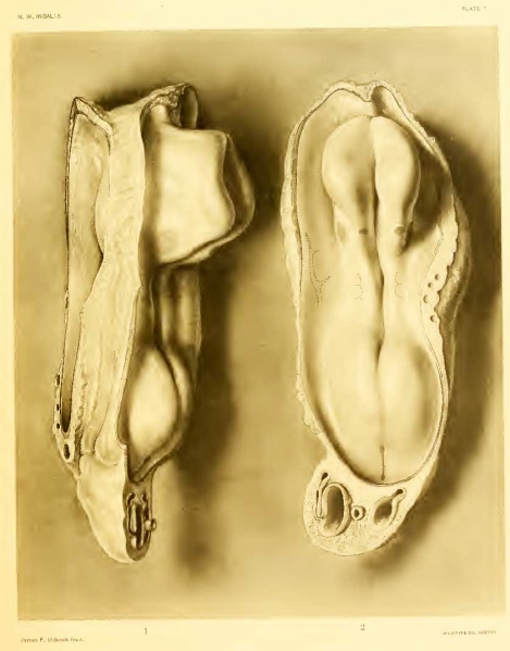
Plate 1
Fig. 1.
- Left lateral view of model of embryonic body viewed in the plane of section.
- X 100. Yolk-sac, amnion, and body-stalk have been cut away.
Fig. 2.
- Dorsal view of the same model.
Both views show open neural groove. Otic plates and V ganglia appear as darker, rounded areas, the former larger and farther lateral, in the region of the rhombencephalon.
Outlines of the underlying somites and in figure 2 the left umbilical vein.
At the posterior end of the body the broad, dark line indicates the primitive-streak region with the primitive node at its anterior end and the cloacal membrane at the posterior end.
Projecting from the cut surface of the body-stalk are the two umbilical arteries, with their branches to form the ventral venous plexus (cf . figs. 3 and 4) ; between the arteries is the allantois.
In figure 1, first ectodermic pocket in front of left otic plate, opposite midbrain flexure.
- Embryo at Segmentation: Figure A | Plate 1 | Plate 2 | Plate 3 | Plate 4 | Plate 5 | Carnegie stage 9 | Carnegie Embryo 1878
Reference
Ingalls NW. A human embryo at the beginning of segmentation, with special reference to the vascular system. (1920) Contrib. Embryol., Carnegie Inst. Wash. Publ. 274, 11: 61-90.
Cite this page: Hill, M.A. (2024, April 19) Embryology Ingalls1920plate01.jpg. Retrieved from https://embryology.med.unsw.edu.au/embryology/index.php/File:Ingalls1920plate01.jpg
- © Dr Mark Hill 2024, UNSW Embryology ISBN: 978 0 7334 2609 4 - UNSW CRICOS Provider Code No. 00098G
| Historic Disclaimer - information about historic embryology pages |
|---|
| Pages where the terms "Historic" (textbooks, papers, people, recommendations) appear on this site, and sections within pages where this disclaimer appears, indicate that the content and scientific understanding are specific to the time of publication. This means that while some scientific descriptions are still accurate, the terminology and interpretation of the developmental mechanisms reflect the understanding at the time of original publication and those of the preceding periods, these terms, interpretations and recommendations may not reflect our current scientific understanding. (More? Embryology History | Historic Embryology Papers) |
File history
Click on a date/time to view the file as it appeared at that time.
| Date/Time | Thumbnail | Dimensions | User | Comment | |
|---|---|---|---|---|---|
| current | 18:04, 30 January 2012 |  | 837 × 1,069 (53 KB) | S8600021 (talk | contribs) | {{Ingalls1920}} {{Historic Disclaimer}} |
You cannot overwrite this file.
File usage
The following 4 pages use this file:
