File:Human spermatozoa nucleus EM03.jpg
Human_spermatozoa_nucleus_EM03.jpg (600 × 475 pixels, file size: 44 KB, MIME type: image/jpeg)
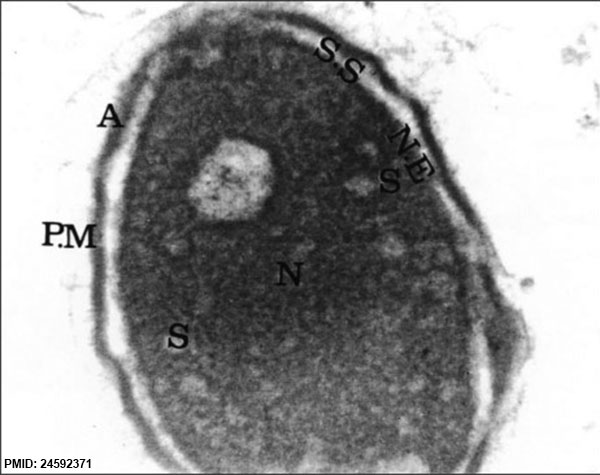
Human Spermatozoa Nucleus EM
Electron micrograph image of a cross section of a abnormal spermatozoa head, nucleus has a less uniform and less condensed chromatin.
Longitudinal section of an abnormal sperm head belonging to the group with high fluorescence rate. Nucleus is observed with insufficiently condensed chromatin and irregular sperm plasma membrane (PM), acrosome (A) and nuclear envelope (NE). Acrosome (A) and subacrosome space (SS) do not have uniform thicknesses in all places (magnification of ×20000)
- Human Spermatozoa EM: Image - cap-phase spermatid | Image - elongated spermatid | Image - spermatid | Image - spermatozoa | Image - normal nucleus | Image - nucleus | Image - abnormal nucleus | Spermatozoa Development | Category:Electron Micrograph
Reference
<pubmed>24592371</pubmed>| Adv Biomed Res.
Copyright
© 2014 Iranpour
This is an open-access article distributed under the terms of the Creative Commons Attribution License (http://creativecommons.org/licenses/by/2.0), which permits unrestricted use, distribution, and reproduction in any medium, provided the original work is properly cited.
Figure 3 http://www.advbiores.net/viewimage.asp?img=AdvBiomedRes_2014_3_1_24_124666_f3.jpg Image adjusted in size, contrast and labelling.
File history
Click on a date/time to view the file as it appeared at that time.
| Date/Time | Thumbnail | Dimensions | User | Comment | |
|---|---|---|---|---|---|
| current | 09:39, 17 September 2014 | | 600 × 475 (44 KB) | Z8600021 (talk | contribs) | ==Human Spermatozoa Nucleus EM== Electron micrograph image of a cross section of a normal spermatozoa head, nucleus has a less uniform and less condensed chromatin. Nucleus belonging to group with high fluorescence rate observed with insufficiently c... |
You cannot overwrite this file.
File usage
The following page uses this file: