File:Human spermatid EM01.jpg

Original file (1,000 × 762 pixels, file size: 162 KB, MIME type: image/jpeg)
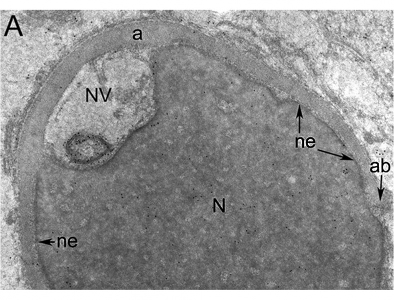
Human Cap-phase Spermatid Electron Micrograph
Cap-phase spermatid (labelling with anti-SPANXa/d antibodies) staining was not observed associated with the nuclear envelope (ne) underlying the developing acrosome (a). However, gold particles (5 nm) were present on membrane whorls lying within a nuclear vacuole (NV) that opened into the subacrosomal space.
A × 20,000
- Human Spermatozoa EM: Image - cap-phase spermatid | Image - elongated spermatid | Image - spermatid | Image - spermatozoa | Image - normal nucleus | Image - nucleus | Image - abnormal nucleus | Spermatozoa Development | Category:Electron Micrograph
- Spermatozoa Images: Spermatozoa BF | Spermatozoon BF | Spermatozoon EM | Spermatozoon EM | Historic drawing | Category:Spermatozoa | Spermatozoa Development | Testis Development
Reference
<pubmed>17012309</pubmed>| Mol Hum Reprod.
Copyright
© The Author 2006. Published by Oxford University Press on behalf of the European Society of Human Reproduction and Embryology. All rights reserved. For Permissions, please email: journals.permissions@oxfordjournals.org
The online version of this article has been published under an open access model. Users are entitled to use, reproduce, disseminate, or display the open access version of this article for non-commercial purposes provided that: the original authorship is properly and fully attributed; the Journal and Oxford University Press are attributed as the original place of publication with the correct citation details given; if an article is subsequently reproduced or disseminated not in its entirety but only in part or as a derivative work this must be clearly indicated. For commercial re-use, please contact journals.permissions@oxfordjournals.org
Original file name: Figure 8. http://molehr.oxfordjournals.org/content/12/11/703/F8.expansion.html Panel A cropped and resized from original figure.
File history
Click on a date/time to view the file as it appeared at that time.
| Date/Time | Thumbnail | Dimensions | User | Comment | |
|---|---|---|---|---|---|
| current | 10:14, 17 September 2014 | | 1,000 × 762 (162 KB) | Z8600021 (talk | contribs) | ==Human Spermatid Electron Micrograph== Electron micrographs of human spermatids following post-embedding labelling with anti-SPANXa/d antibodies. * '''A''' - cap-phase spermatid staining was not observed associated with the nuclear envelope (ne) un... |
You cannot overwrite this file.
File usage
The following page uses this file: