File:Human embryo skin 24 week EGA.jpg
Original file (596 × 939 pixels, file size: 165 KB, MIME type: image/jpeg)
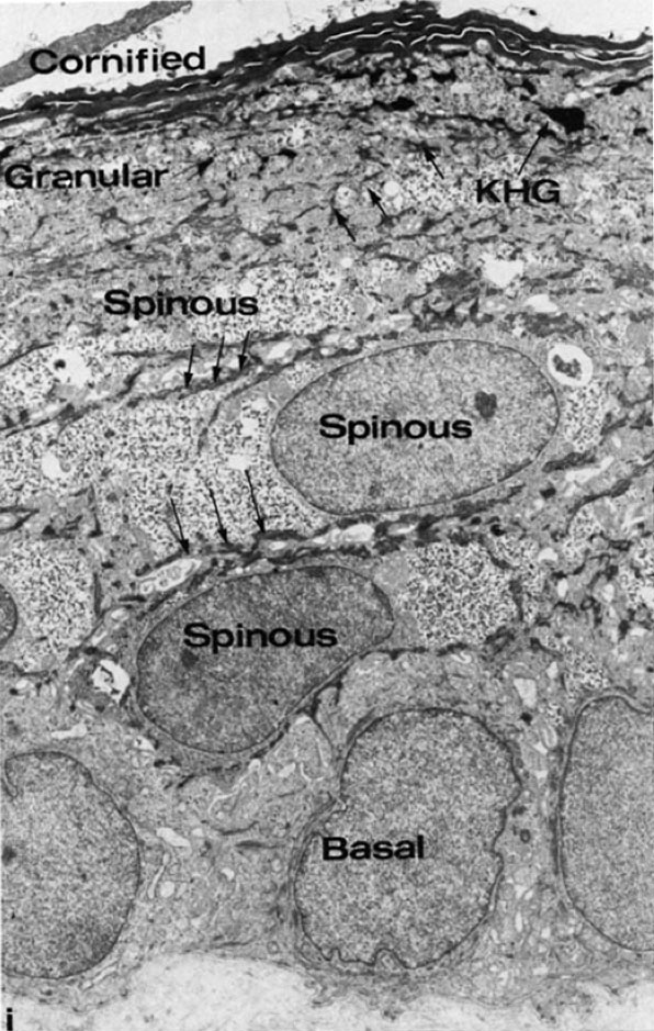
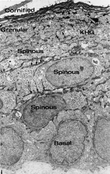
Keratinized Fetal Epidermis (about 24 week EGA)
Transmission electron micrograph (TEM) of full thickness keratinized epidermis. Note the presence of abundant glycogen in all epidermal layers, the peripherally organized bundles of keratin filaments (arrows), and small keratohyalin granules (KHG) in one or two layers of granular cells.
- 24 weeks Estimated Gestational Age = about 22 weeks Fertilization Age
- Magnification x 4,625. (cropped from figure 4, contrast adjusted)
- Links: Epidermis 8-9 week EGA | Epidermis desmosomes 8-9 week EGA | Epidermis 9-11 week EGA | Epidermis 24 week EGA | Integumentary System Development
Reference
<pubmed>2413039</pubmed>| PMC2113922
Copyright
Rockefeller University Press - Copyright Policy This article is distributed under the terms of an Attribution–Noncommercial–Share Alike–No Mirror Sites license for the first six months after the publication date (see http://www.jcb.org/misc/terms.shtml). After six months it is available under a Creative Commons License (Attribution–Noncommercial–Share Alike 4.0 Unported license, as described at https://creativecommons.org/licenses/by-nc-sa/4.0/ ). (More? Help:Copyright Tutorial)
File history
Click on a date/time to view the file as it appeared at that time.
| Date/Time | Thumbnail | Dimensions | User | Comment | |
|---|---|---|---|---|---|
| current | 22:54, 28 September 2011 | | 596 × 939 (165 KB) | S8600021 (talk | contribs) | ==Keratinized Fetal Epidermis_(about 24 week EGA)== Transmission electron micrograph of full thickness keratinized epidermis. Note the presence of abundant glycogen in all epidermal layers, the peripherally organized bundles of keratin filaments (arrow |
You cannot overwrite this file.
File usage
The following 4 pages use this file: