File:Human- fetal week 10 head A1.jpg
From Embryology

Size of this preview: 661 × 599 pixels. Other resolution: 1,200 × 1,088 pixels.
Original file (1,200 × 1,088 pixels, file size: 159 KB, MIME type: image/jpeg)
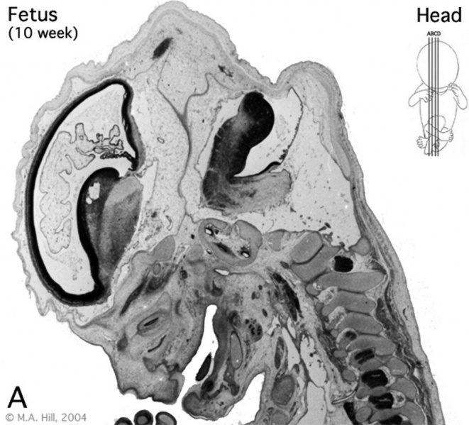
Human Fetus
female, 10 week, 40 mm CRL, early fetal, sagittal section, pelvic region
This stage of development is after the embryonic period (up to week 8) but still only 2 weeks into early fetal development.
Section A is the most sagittal (lateral towards right) of all sections, plane B, C and D move towards the midline.
Original file name: H10wkHeadA.jpg
Related Images
Fetus (week 10) Planes A (most lateral), B (lateral), C (medial) and D (midline) from lateral towards the midline.
- Human Fetus - most lateral | lateral | medial | midline
- Head - most lateral | lateral | medial | midline
- Cerebellum - most lateral | lateral | medial | midline
- Urogenital Unlabelled - most lateral | lateral | medial | midline
- Urogenital Labelled - most lateral | lateral | medial | midline
- Large Images - midline
- Image Source: UNSW Embryology, no reproduction without permission.
Human- fetal week 10 head A.jpg
H10wkHeadAlarge.jpg
File history
Click on a date/time to view the file as it appeared at that time.
| Date/Time | Thumbnail | Dimensions | User | Comment | |
|---|---|---|---|---|---|
| current | 06:58, 7 October 2010 | | 1,200 × 1,088 (159 KB) | S8600021 (talk | contribs) | ==Human Fetus== female, 10 week, 40 mm CRL, early fetal, sagittal section, pelvic region This stage of development is after the embryonic period (up to week 8) but still only 2 weeks into early fetal development. Section A is the most sagittal (lateral |
You cannot overwrite this file.
File usage
There are no pages that use this file.