File:Historic-lungs.jpg
From Embryology
No higher resolution available.
Historic-lungs.jpg (600 × 493 pixels, file size: 79 KB, MIME type: image/jpeg)
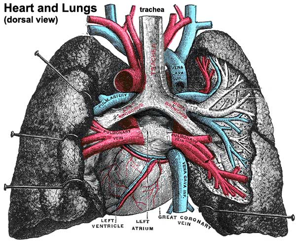
Adult Heart and Lungs Anatomy
Historic drawing of the lungs showing dorsal view and anatomical size and position with respect to the heart.
- Each lung - is conical in shape, and presents for examination an apex, a base, three borders, and two surfaces.
- Left lung - is divided into two lobes, an upper and a lower, by an interlobular fissure.
- Right lung - is divided into three lobes, superior, middle, and inferior, by two interlobular fissures.
- Azygos lobe - the right lung upper lobe expands either side of the posterior cardinal. Common condition (0.5% of population) there is also some course variability of the phrenic nerve in the presence of an azygos lobe.
File history
Click on a date/time to view the file as it appeared at that time.
| Date/Time | Thumbnail | Dimensions | User | Comment | |
|---|---|---|---|---|---|
| current | 14:35, 24 August 2009 | | 600 × 493 (79 KB) | MarkHill (talk | contribs) | Historic drawing of the lungs showing dorsal view and anatomical size and position with respect to the heart. Category:Respiratory Category:Historic Category:Gray's 1918 Anatomy |
You cannot overwrite this file.
File usage
The following 3 pages use this file: