File:Histology-fetal liver HEx40.jpg

Original file (1,000 × 800 pixels, file size: 281 KB, MIME type: image/jpeg)
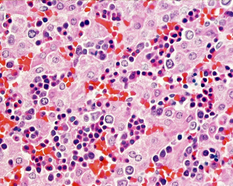
Fetal Liver
This histology section of the fetal liver shows the prresence of large numbers of red blood cells (erythrocytes) and blood stem cells. (Stain - Haematoxylin Eosin)
Note also the presence of nucleated erythrocytes that do not occur in adult blood.
liver fetal human accessory digestive glands
- Links: Liver Histology | Liver Development | Blood Development
Links: Histology | Histology Stains | Blue Histology images copyright Lutz Slomianka 1998-2009. The literary and artistic works on the original Blue Histology website may be reproduced, adapted, published and distributed for non-commercial purposes. See also the page Histology Stains.
Cite this page: Hill, M.A. (2024, April 24) Embryology Histology-fetal liver HEx40.jpg. Retrieved from https://embryology.med.unsw.edu.au/embryology/index.php/File:Histology-fetal_liver_HEx40.jpg
- © Dr Mark Hill 2024, UNSW Embryology ISBN: 978 0 7334 2609 4 - UNSW CRICOS Provider Code No. 00098G
Original file name: lif40he.jpg
File history
Click on a date/time to view the file as it appeared at that time.
| Date/Time | Thumbnail | Dimensions | User | Comment | |
|---|---|---|---|---|---|
| current | 07:51, 23 August 2010 | | 1,000 × 800 (281 KB) | S8600021 (talk | contribs) | ==Fetal Liver== Original file name: lif40he.jpg liver fetal human HE accessory digestive glands {{Template:Blue Histology}} Category:Histology Category:Liver Category:Gastrointestinal Tract |
You cannot overwrite this file.
File usage
The following 3 pages use this file: