File:Heart chicken embryo stage 25.jpg

Original file (1,000 × 1,154 pixels, file size: 219 KB, MIME type: image/jpeg)
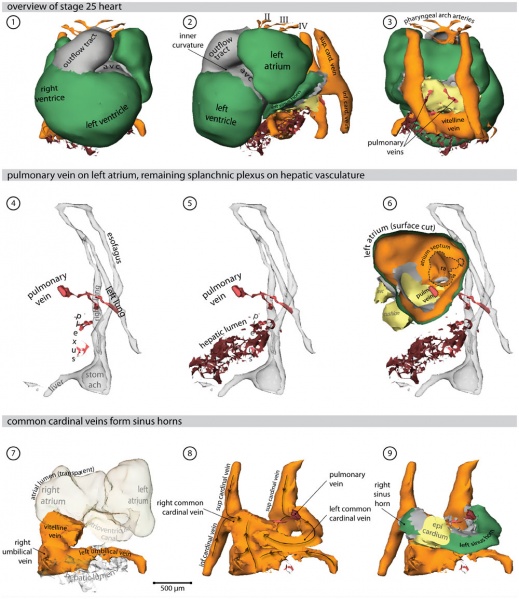
Chicken Heart (stage 25 embryo)
- Ventral view of the heart. Cx40 positive working myocardium is shown in green, primary myocardium is depicted in grey.
- Left lateral view of the heart; the splanchnic plexus is shown in red and hepatic lumen in brown.
- Dorsal view of the heart.
- Left lateral view of the endoderm (transparent grey) in relation to the pulmonary vein and the splanchnic plexus.
- The remaining plexus attaches to the hepatic lumen.
- Left lateral view; the surface of the left atrium is cut, showing the atrial septum and the entrance of the pulmonary vein.
- Systemic venous returns (orange) with respect to the atrial lumen (transparent).
- Direction of blood-flow.
- The common cardinal veins entirely ensleeved by myocardium. Most of this myocardium now expresses Cx40, with the exception of the forming sinus node.
Abbreviations
- I, II, III - first, second & third pharyngeal arch artery
- avc - atrioventricular canal
- Links: Image - Heart 3D reconstruction | Image - Stage 12 Heart | Image - Stage 16 Heart | Image - Stage 21 Heart | Image - Stage 25 Heart | Hamburger Hamilton Stages | Cardiovascular System Development | Chicken Development
Online Editor - Note image is labeled stage 22, but paper text refers to stage 21.
Reference
<pubmed>21779373</pubmed>| PLoS One
Copyright
© 2011 van den Berg, Moorman. This is an open-access article distributed under the terms of the Creative Commons Attribution License, which permits unrestricted use, distribution, and reproduction in any medium, provided the original author and source are credited.
Original image name: Figure 6. Journal.pone.0022055.g006.jpg http://www.ncbi.nlm.nih.gov/pmc/articles/PMC3133620/figure/pone-0022055-g006/
Cite this page: Hill, M.A. (2024, April 17) Embryology Heart chicken embryo stage 25.jpg. Retrieved from https://embryology.med.unsw.edu.au/embryology/index.php/File:Heart_chicken_embryo_stage_25.jpg
- © Dr Mark Hill 2024, UNSW Embryology ISBN: 978 0 7334 2609 4 - UNSW CRICOS Provider Code No. 00098G
File history
Click on a date/time to view the file as it appeared at that time.
| Date/Time | Thumbnail | Dimensions | User | Comment | |
|---|---|---|---|---|---|
| current | 10:50, 27 August 2011 | | 1,000 × 1,154 (219 KB) | S8600021 (talk | contribs) | ==Chicken Heart (stage 25 embryo)== # Ventral view of the heart. Cx40 positive working myocardium is shown in green, primary myocardium is depicted in grey. # Left lateral view of the heart; the splanchnic plexus is shown in red and hepatic lumen in bro |
You cannot overwrite this file.
File usage
The following 2 pages use this file: