File:Heart chicken embryo stage 21.jpg
Original file (1,000 × 1,197 pixels, file size: 207 KB, MIME type: image/jpeg)
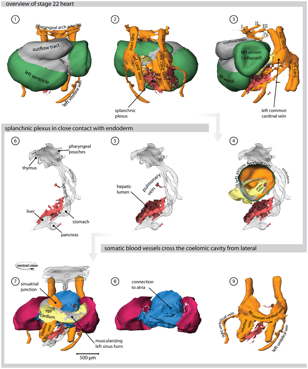
Chicken Heart (stage 21 embryo)
- Ventral view of the heart. Cx40 positive working myocardium is shown in green, primary myocardium is depicted in grey. # Dorsal view; the splanchnic plexus is shown in light red and hepatic lumen in brown.
- Left lateral view.
- Left lateral view; the surface of the left atrium is cut, showing the atrial septum and the entrance of the pulmonary vein. The endoderm is transparent.
- The splanchnic plexus is interwoven with the hepatic lumen
- Within the endoderm several organs are developing.
- Ventral view showing the sinus venosus region with respect to the splanchnic (blue) and somatic (red) mesoderm
- Solitary depiction of the splanchnic and the somatic mesoderm. The lateral mesocardium is indicated by a hatched line.
- The venous systems of the cardiac inflow.
Abbreviations
- I, II, III - first, second & third pharyngeal arch artery
- la - left atrium
- ra - right atrium
- avc - atrioventricular canal
- sup. card. - superior cardinal vein
- inf. card. - inferior cardinal vein
- pulm. vein - pulmonary vein
- Links: Image - Heart 3D reconstruction | Image - Stage 12 Heart | Image - Stage 16 Heart | Image - Stage 21 Heart | Image - Stage 25 Heart | Hamburger Hamilton Stages | Cardiovascular System Development | Chicken Development
Note image is labeled stage 22, but paper text refers to stage 21.
Reference
<pubmed>21779373</pubmed>| PLoS One
Copyright
© 2011 van den Berg, Moorman. This is an open-access article distributed under the terms of the Creative Commons Attribution License, which permits unrestricted use, distribution, and reproduction in any medium, provided the original author and source are credited.
Original image name: Figure 4. Journal.pone.0022055.g005.jpg http://www.ncbi.nlm.nih.gov/pmc/articles/PMC3133620/figure/pone-0022055-g005/
Cite this page: Hill, M.A. (2024, April 16) Embryology Heart chicken embryo stage 21.jpg. Retrieved from https://embryology.med.unsw.edu.au/embryology/index.php/File:Heart_chicken_embryo_stage_21.jpg
- © Dr Mark Hill 2024, UNSW Embryology ISBN: 978 0 7334 2609 4 - UNSW CRICOS Provider Code No. 00098G
File history
Click on a date/time to view the file as it appeared at that time.
| Date/Time | Thumbnail | Dimensions | User | Comment | |
|---|---|---|---|---|---|
| current | 10:45, 27 August 2011 | | 1,000 × 1,197 (207 KB) | S8600021 (talk | contribs) | ==Chicken Heart (stage 21 embryo)== # Ventral view of the heart. Cx40 positive working myocardium is shown in green, primary myocardium is depicted in grey. # Dorsal view; the splanchnic plexus is shown in light red and hepatic lumen in brown. # Left lat |
You cannot overwrite this file.
File usage
The following 2 pages use this file: