File:Heart Tube Segments.jpg

Original file (1,082 × 771 pixels, file size: 63 KB, MIME type: image/jpeg)
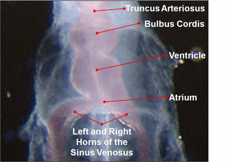
Early Human Heart Tube
Ventral view Week 4, Carnegie stage 10
As the tubular heart grows it develops dilations and constrictions which form the truncus arteriosus, bulbus cordis, primitive ventricle, primitive atrium and sinus venosus.
| Begin Intermediate: | Primordial Heart Tube | Heart Tube Looping | Atrial Ventricular Septation | Outflow Tract | Heart Valves | Cardiac Abnormalities | Vascular Overview |
Image Source: Scanning electron micrographs of the Carnegie stages of the early human embryos are reproduced with the permission of Prof Kathy Sulik, from embryos collected by Dr. Vekemans and Tania Attié-Bitach. Images are for educational purposes only and cannot be reproduced electronically or in writing without permission.
Cite this page: Hill, M.A. (2024, April 19) Embryology Heart Tube Segments.jpg. Retrieved from https://embryology.med.unsw.edu.au/embryology/index.php/File:Heart_Tube_Segments.jpg
- © Dr Mark Hill 2024, UNSW Embryology ISBN: 978 0 7334 2609 4 - UNSW CRICOS Provider Code No. 00098G
File history
Click on a date/time to view the file as it appeared at that time.
| Date/Time | Thumbnail | Dimensions | User | Comment | |
|---|---|---|---|---|---|
| current | 10:42, 14 March 2010 | | 1,082 × 771 (63 KB) | Z3212774 (talk | contribs) | category:Heart ILP {{Template:SEM}} As the tubular heart grows it develops dilations and constrictions which form the truncus arteriosus, bulbus cordis, primitive ventricle, primitive atrium and sinus venosus. |
You cannot overwrite this file.
File usage
The following file is a duplicate of this file (more details):
The following 19 pages use this file:
- 2010 Lab 4
- 2011 Lab 4
- ANAT2341 Lab 11 - Heart
- ANAT2341 Lab 4 - Early Cardiovascular Development
- B
- BGDA Practical 7 - Week 4
- Cardiovascular - Arterial Development
- Cardiovascular - Venous Development
- Cardiovascular System - Circulation Development
- Cardiovascular System - Coronary Circulation Development
- Cardiovascular System - Truncus Arteriosus
- Cardiovascular System Development
- Carnegie stage 10
- Fetal ECHO Meeting 2012
- Human Embryo SEM
- Intermediate - Primordial Heart Tube
- Lecture - Early Vascular Development
- Lecture - Heart Development
- RPAH Cardiac Embryology 2014