File:Hair follicle development 02.jpg

Original file (800 × 616 pixels, file size: 75 KB, MIME type: image/jpeg)
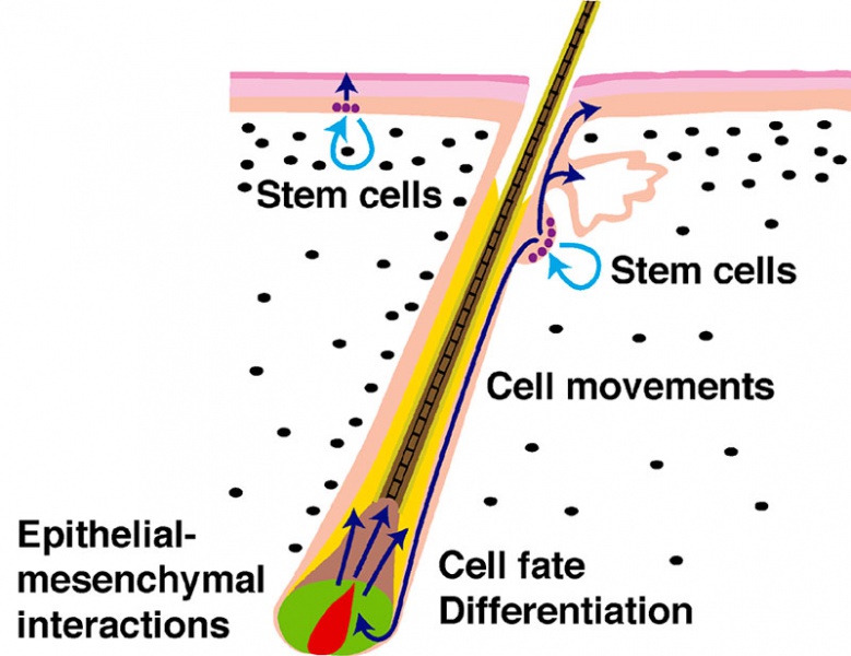
Hair Follicle Development cartoon
Postnatal skin showing a hair follicle in the growth phase. Biological processes occurring in the skin are listed and stem cell locations are indicated.
| Legend | |
|---|---|
|
|
- Hair Development: Follicle stages | Follicle Stem Cells | Skin structure | Mouse follicle histology | Hair Development
Reference
<pubmed>16277556</pubmed>| PLoS Biol.
Copyright
© 2005 Sarah E. Millar. This is an open-access article distributed under the terms of the Creative Commons Attribution License, which permits unrestricted use, distribution, and reproduction in any medium, provided the original author and source are credited.
Citation: Millar SE (2005) An Ideal Society? Neighbors of Diverse Origins Interact to Create and Maintain Complex Mini-Organs in the Skin. PLoS Biol 3(11): e372. doi:10.1371/journal.pbio.0030372
Image cropped and resized from Figure 1 panel B.
File history
Click on a date/time to view the file as it appeared at that time.
| Date/Time | Thumbnail | Dimensions | User | Comment | |
|---|---|---|---|---|---|
| current | 11:00, 23 March 2014 | | 800 × 616 (75 KB) | Z8600021 (talk | contribs) |
You cannot overwrite this file.
File usage
The following 2 pages use this file: