File:Gray1119.jpg
From Embryology
Size of this preview: 520 × 599 pixels. Other resolution: 700 × 807 pixels.
Original file (700 × 807 pixels, file size: 115 KB, MIME type: image/jpeg)
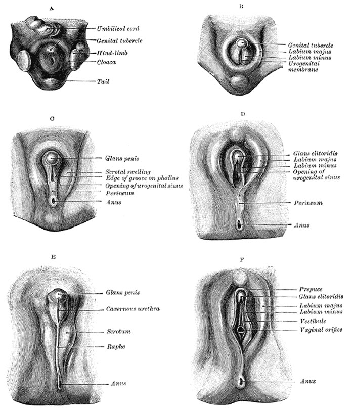
Stages in the Development of the External Sexual Organs in the Male and Female
- Drawn from the Ecker-Ziegler models.
| Historic Disclaimer - information about historic embryology pages |
|---|
 |
The External Organs of Generation
- cloacal membrane, composed of ectoderm and endoderm (entoderm), originally reaches from the umbilicus to the tail
- mesoderm extends to the mid-ventral line for some distance behind the umbilicus
- forms the lower part of the abdominal wall
- ends below in a prominent swelling, the cloacal tubercle
- Behind the tubercle the urogenital part of the cloacal membrane separates the ingrowing sheets of mesoderm
- The first rudiment of the penis (or clitoris) is a structure termed the phallus
- derived from the phallic portion of the cloaca which has extended on to the end and sides of the under surface of the cloacal tubercle
- The terminal part of the phallus representing the future glans becomes solid
- the remainder, which is hollow, is converted into a longitudinal groove by the absorption of the urogenital membrane
Female
- a deep groove forms around the phallus and separates it from the rest of the cloacal tubercle, which is now termed the genital tubercle.
- sides of the genital tubercle grow backward as the genital swellings, which ultimately form the labia majora
- tubercle itself becomes the mons pubis
- labia minora arise by the continued growth of the lips of the groove on the under surface of the phallus
- remainder of the phallus forms the clitoris
Male
- early changes are similar
- pelvic portion of the cloaca undergoes much greater development, pushing before it the phallic portion
- genital swellings extend around between the pelvic portion and the anus, and form a scrotal area
- during the changes associated with the descent of the testes this area is drawn out to form the scrotal sac
- penis is developed from the phallus
- urogenital membrane undergoes absorption, forming a channel on the under surface of the phallus; this channel extends only as far forward as the corona glandis
(Text modified from Gray's Anatomy)
entoderm - is a historic term for endoderm.
- Gray's Images: Development | Lymphatic | Neural | Vision | Hearing | Somatosensory | Integumentary | Respiratory | Gastrointestinal | Urogenital | Endocrine | Surface Anatomy | iBook | Historic Disclaimer
| Historic Disclaimer - information about historic embryology pages |
|---|
|
| iBook - Gray's Embryology | |
|---|---|

|
|
Reference
Gray H. Anatomy of the human body. (1918) Philadelphia: Lea & Febiger.
Cite this page: Hill, M.A. (2024, April 18) Embryology Gray1119.jpg. Retrieved from https://embryology.med.unsw.edu.au/embryology/index.php/File:Gray1119.jpg
- © Dr Mark Hill 2024, UNSW Embryology ISBN: 978 0 7334 2609 4 - UNSW CRICOS Provider Code No. 00098G
File history
Click on a date/time to view the file as it appeared at that time.
| Date/Time | Thumbnail | Dimensions | User | Comment | |
|---|---|---|---|---|---|
| current | 08:06, 28 May 2011 | | 700 × 807 (115 KB) | S8600021 (talk | contribs) | ==Stages in the development of the external sexual organs in the male and female== * Drawn from the Ecker-Ziegler models. {{Historic Disclaimer}} ===The External Organs of Generation === * The cloacal membrane, composed of ectoderm and endoderm (entoder |
You cannot overwrite this file.