File:Gray1030.jpg
Original file (481 × 800 pixels, file size: 122 KB, MIME type: image/jpeg)
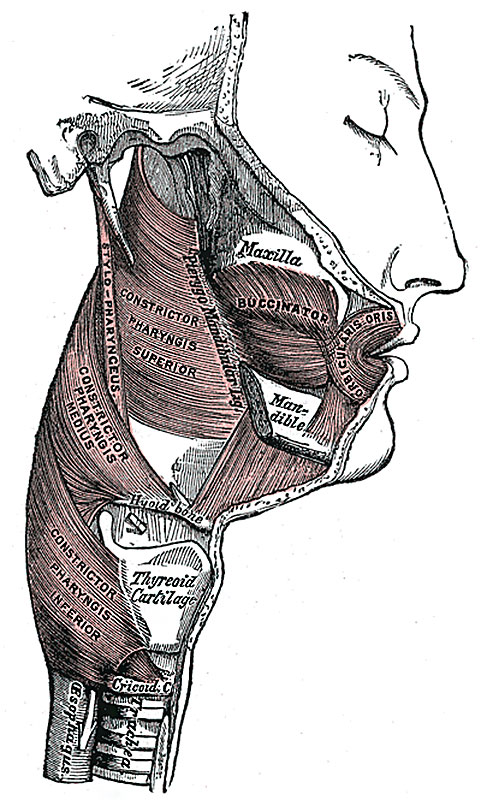
Fig. 1030 Muscles of the Pharynx and Cheek
The muscles of the pharynx are:
- Constrictor superior - Pharyngopalatinus.
- Constrictor medius - Salpingopharyngeus.
- Constrictor inferior - Stylopharyngeus.
Constrictor pharynges superior
(Superior constrictor) (Fig. 1030, 1031) is a quadrilateral muscle, thinner and paler than the other two. It arises from the lower third of the posterior margin of the medial pterygoid plate and its hamulus, from the pterygomandibular raphé, from the alveolar process of the mandible above the posterior end of the mylohyoid line, and by a few fibers from the side of the tongue. The fibers curve backward to be inserted into the median raphé, being also prolonged by means of an aponeurosis to the pharyngeal spine on the basilar part of the occipital bone. The superior fibers arch beneath the Levator veli palatini and the auditory tube. The interval between the upper border of the muscle and the base of the skull is closed by the pharyngeal aponeurosis, and is known as the sinus of Morgagni.
Constrictor pharynges medius
(Middle constrictor) (Figs. 1030, 1031) is a fanshaped muscle, smaller than the preceding. It arises from the whole length of the upper border of the greater cornu of the hyoid bone, from the lesser cornu, and from the stylohyoid ligament. The fibers diverge from their origin: the lower ones descend beneath the Constrictor inferior, the middle fibers pass transversely, and the upper fibers ascend and overlap the Constrictor superior. It is inserted into the posterior median fibrous raphé, blending in the middle line with the muscle of the opposite side.
Constrictor pharynges inferior
(Inferior constrictor) (Figs. 1030, 1031), the thickest of the three constrictors, arises from the sides of the cricoid and thyroid cartilage. From the cricoid cartilage it arises in the interval between the Cricothyreoideus in front, and the articular facet for the inferior cornu of the thyroid cartilage behind. On the thyroid cartilage it arises from the oblique line on the side of the lamina, from the surface behind this nearly as far as the posterior border and from the inferior cornu. From these origins the fibers spread backward and medialward to be inserted with the muscle of the opposite side into the fibrous raphé in the posterior median line of the pharynx. The inferior fibers are horizontal and continuous with the circular fibers of the esophagus; the rest ascend, increasing in obliquity, and overlap the Constrictor medius.
- Gray's Images: Development | Lymphatic | Neural | Vision | Hearing | Somatosensory | Integumentary | Respiratory | Gastrointestinal | Urogenital | Endocrine | Surface Anatomy | iBook | Historic Disclaimer
| Historic Disclaimer - information about historic embryology pages |
|---|
 |
| iBook - Gray's Embryology | |
|---|---|

|
|
Reference
Gray H. Anatomy of the human body. (1918) Philadelphia: Lea & Febiger.
Cite this page: Hill, M.A. (2024, April 25) Embryology Gray1030.jpg. Retrieved from https://embryology.med.unsw.edu.au/embryology/index.php/File:Gray1030.jpg
- © Dr Mark Hill 2024, UNSW Embryology ISBN: 978 0 7334 2609 4 - UNSW CRICOS Provider Code No. 00098G
File history
Click on a date/time to view the file as it appeared at that time.
| Date/Time | Thumbnail | Dimensions | User | Comment | |
|---|---|---|---|---|---|
| current | 14:22, 8 September 2014 | | 481 × 800 (122 KB) | Z8600021 (talk | contribs) | :'''Links:''' Tongue Development | Head Development | Musculoskeletal System Development {{Gray Anatomy}} Category:Tongue Category:Head Category:Skeletal MuscleCategory:Cartoon |
You cannot overwrite this file.
File usage
The following 2 pages use this file: