File:Gray0991.jpg
Gray0991.jpg (469 × 400 pixels, file size: 33 KB, MIME type: image/jpeg)
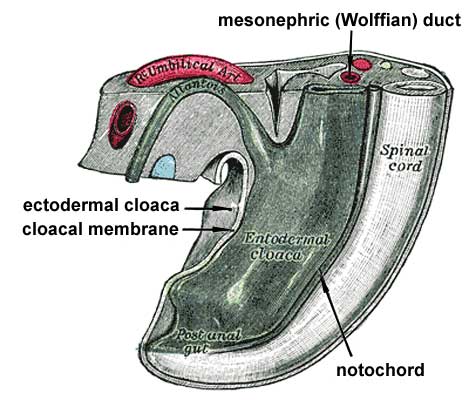
Week 3 - Tail end of human embryo from fifteen to eighteen days old
From model by Franz Keibel (1861-1929)
The Rectum and Anal Canal - The hind-gut is at first prolonged backward into the body-stalk as the tube of the allantois; but, with the growth and flexure of the tail-end of the embryo, the body-stalk, with its contained allantoic tube, is carried forward to the ventral aspect of the body, and consequently a bend is formed at the junction of the hind-gut and allantois. This bend becomes dilated into a pouch, which constitutes the entodermal cloaca; into its dorsal part the hind-gut opens, and from its ventral part the allantois passes forward. At a later stage the Wolffian and Müllerian ducts open into its ventral portion. The cloaca is, for a time, shut off from the anterior by a membrane, the cloacal membrane, formed by the apposition of the ectoderm and entoderm, and reaching, at first, as far forward as the future umbilicus. Behind the umbilicus, however, the mesoderm subsequently extends to form the lower part of the abdominal wall and symphysis pubis. By the growth of the surrounding tissues the cloacal membrane comes to lie at the bottom of a depression, which is lined by ectoderm and named the ectodermal cloaca (Fig. 991).
- Links: Image 987a | Image 987b | Image - Early Week 4 | Image - Late Week 4 | Gastrointestinal Tract Development | Endoderm
- Gray's Images: Development | Lymphatic | Neural | Vision | Hearing | Somatosensory | Integumentary | Respiratory | Gastrointestinal | Urogenital | Endocrine | Surface Anatomy | iBook | Historic Disclaimer
| Historic Disclaimer - information about historic embryology pages |
|---|
 |
| iBook - Gray's Embryology | |
|---|---|

|
|
Reference
Gray H. Anatomy of the human body. (1918) Philadelphia: Lea & Febiger.
Cite this page: Hill, M.A. (2024, April 23) Embryology Gray0991.jpg. Retrieved from https://embryology.med.unsw.edu.au/embryology/index.php/File:Gray0991.jpg
- © Dr Mark Hill 2024, UNSW Embryology ISBN: 978 0 7334 2609 4 - UNSW CRICOS Provider Code No. 00098G
File history
Click on a date/time to view the file as it appeared at that time.
| Date/Time | Thumbnail | Dimensions | User | Comment | |
|---|---|---|---|---|---|
| current | 15:47, 23 August 2009 | | 469 × 400 (33 KB) | S8600021 (talk | contribs) |
You cannot overwrite this file.
File usage
The following 9 pages use this file:
- 2009 Lecture 9
- 2010 Lecture 9
- Anatomy of the Human Body by Henry Gray
- BGDB Sexual Differentiation - Late Embryo
- Gastrointestinal Tract - Mouth Development
- Gastrointestinal Tract - Oesophagus Development
- Gastrointestinal Tract Development
- Lecture - Gastrointestinal Development 2013
- Talk:2010 Lecture 9