File:Gray0979.jpg
Gray0979.jpg (500 × 446 pixels, file size: 56 KB, MIME type: image/jpeg)
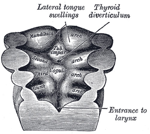
Floor of pharynx of human embryo about twenty-six days old
(From model by Peters.)
The Tongue
(Figs. 979 to 981) The tongue is developed in the floor of the pharynx, and consists of an anterior or buccal and a posterior or pharyngeal part which are separated in the adult by the V-shaped sulcus terminalis. During the third week there appears, immediately behind the ventral ends of the two halves of the mandibular arch, a rounded swelling named the tuberculum impar, which was described by His as undergoing enlargement to form the buccal part of the tongue. More recent researches, however, show that this part of the tongue is mainly, if not entirely, developed from a pair of lateral swellings which rise from the inner surface of the mandibular arch and meet in the middle line.
The tuberculum impar is said to form the central part of the tongue immediately in front of the foramen cecum, but Hammar insists that it is purely a transitory structure and forms no part of the adult tongue. From the ventral ends of the fourth arch there arises a second and larger elevation, in the center of which is a median groove or furrow. This elevation was named by His the furcula, and is at first separated from the tuberculum impar by a depression, but later by a ridge, the copula, formed by the forward growth and fusion of the ventral ends of the second and third arches.
The posterior or pharyngeal part of the tongue is developed from the copula, which extends forward in the form of a V, so as to embrace between its two limbs the buccal part of the tongue. At the apex of the V a pit-like invagination occurs, to form the thyroid gland, and this depression is represented in the adult by the foramen cecum of the tongue. In the adult the union of the anterior and posterior parts of the tongue is marked by the V-shaped sulcus terminalis, the apex of which is at the foramen cecum, while the two limbs run lateralward and forward, parallel to, but a little behind, the vallate papillæ.
- Links: Tongue Development
- Gray's Images: Development | Lymphatic | Neural | Vision | Hearing | Somatosensory | Integumentary | Respiratory | Gastrointestinal | Urogenital | Endocrine | Surface Anatomy | iBook | Historic Disclaimer
| Historic Disclaimer - information about historic embryology pages |
|---|
 |
| iBook - Gray's Embryology | |
|---|---|

|
|
Reference
Gray H. Anatomy of the human body. (1918) Philadelphia: Lea & Febiger.
Cite this page: Hill, M.A. (2024, April 25) Embryology Gray0979.jpg. Retrieved from https://embryology.med.unsw.edu.au/embryology/index.php/File:Gray0979.jpg
- © Dr Mark Hill 2024, UNSW Embryology ISBN: 978 0 7334 2609 4 - UNSW CRICOS Provider Code No. 00098G
File history
Click on a date/time to view the file as it appeared at that time.
| Date/Time | Thumbnail | Dimensions | User | Comment | |
|---|---|---|---|---|---|
| current | 00:07, 22 August 2012 | | 500 × 446 (56 KB) | Z8600021 (talk | contribs) | {{Gray Anatomy}} Category:Gastrointestinal Tract Category:Human Category:Pharyngeal Arch |
You cannot overwrite this file.
File usage
The following 2 pages use this file: