File:Gray0971.jpg
Gray0971.jpg (800 × 583 pixels, file size: 166 KB, MIME type: image/jpeg)
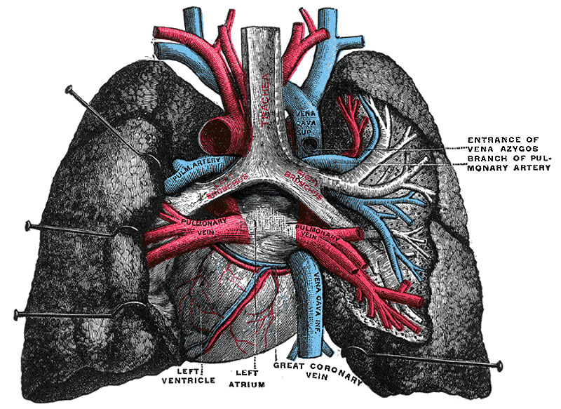
Adult Heart and Lungs Anatomy
Pulmonary vessels, seen in a dorsal view of the heart and lungs. The lungs have been pulled away from the median line, and a part of the right lung has been cut away to display the air-ducts and bloodvessels. (Testut.)
Historic drawing of the adult lungs showing dorsal view and anatomical size and position with respect to the heart.
- Each lung - is conical in shape, and presents for examination an apex, a base, three borders, and two surfaces.
- Left lung - is divided into two lobes, an upper and a lower, by an interlobular fissure.
- Right lung - is divided into three lobes, superior, middle, and inferior, by two interlobular fissures.
- Azygos lobe - the right lung upper lobe expands either side of the posterior cardinal. Common condition (0.5% of population) there is also some course variability of the phrenic nerve in the presence of an azygos lobe.
- Gray's Images: Development | Lymphatic | Neural | Vision | Hearing | Somatosensory | Integumentary | Respiratory | Gastrointestinal | Urogenital | Endocrine | Surface Anatomy | iBook | Historic Disclaimer
| Historic Disclaimer - information about historic embryology pages |
|---|
 |
| iBook - Gray's Embryology | |
|---|---|

|
|
Reference
Gray H. Anatomy of the human body. (1918) Philadelphia: Lea & Febiger.
Cite this page: Hill, M.A. (2024, April 16) Embryology Gray0971.jpg. Retrieved from https://embryology.med.unsw.edu.au/embryology/index.php/File:Gray0971.jpg
- © Dr Mark Hill 2024, UNSW Embryology ISBN: 978 0 7334 2609 4 - UNSW CRICOS Provider Code No. 00098G
File history
Click on a date/time to view the file as it appeared at that time.
| Date/Time | Thumbnail | Dimensions | User | Comment | |
|---|---|---|---|---|---|
| current | 03:12, 17 August 2012 | | 800 × 583 (166 KB) | Z8600021 (talk | contribs) | ==Adult Heart and Lungs Anatomy== Pulmonary vessels, seen in a dorsal view of the heart and lungs. The lungs have been pulled away from the median line, and a part of the right lung has been cut away to display the air-ducts and bloodvessels. (Testut.) |
You cannot overwrite this file.
File usage
The following 4 pages use this file: