File:Gray0955.jpg
Gray0955.jpg (616 × 600 pixels, file size: 91 KB, MIME type: image/jpeg)
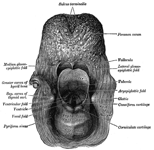
Larynx Entrance
Viewed from behind.
The entrance of the larynx (Fig. 955) is a triangular opening, wide in front, narrow behind, and sloping obliquely downward and backward. It is bounded, in front, by the epiglottis; behind, by the apices of the arytenoid cartilages, the corniculate cartilages, and the interarytenoid notch; and on either side, by a fold of mucous membrane, enclosing ligamentous and muscular fibers, stretched between the side of the epiglottis and the apex of the arytenoid cartilage; this is the aryepiglottic fold, on the posterior part of the margin of which the cuneiform cartilage forms a more or less distinct whitish prominence, the cuneiform tubercle.
Ventricular Folds
(plicœ ventriculares; superior or false vocal cords) Two thick folds of mucous membrane, each enclosing a narrow band of fibrous tissue, the ventricular ligament which is attached in front to the angle of the thyroid cartilage immediately below the attachment of the epiglottis, and behind to the antero-lateral surface of the arytenoid cartilage, a short distance above the vocal process. The lower border of this ligament, enclosed in mucous membrane, forms a free crescentic margin, which constitutes the upper boundary of the ventricle of the larynx.
Vocal Folds
(plicœ vocales; inferior or true vocal cords) Concerned in the production of sound, and enclose two strong bands, named the vocal ligaments (ligamenta vocales; inferior thyroarytenoid). Each ligament consists of a band of yellow elastic tissue, attached in front to the angle of the thyroid cartilage, and behind to the vocal process of the arytenoid. Its lower border is continuous with the thin lateral part of the conus elasticus. Its upper border forms the lower boundary of the ventricle of the larynx. Laterally, the Vocalis muscle lies parallel with it. It is covered medially by mucous membrane, which is extremely thin and closely adherent to its surface.
Ventricle of the Larynx
(ventriculus laryngis [Morgagnii]; laryngeal sinus) A fusiform fossa, situated between the ventricular and vocal folds on either side, and extending nearly their entire length. The fossa is bounded, above, by the free crescentic edge of the ventricular fold; below, by the straight margin of the vocal fold; laterally, by the mucous membrane covering the corresponding Thyreoarytænoideus. The anterior part of the ventricle leads up by a narrow opening into a cecal pouch of mucous membrane of variable size called the appendix.
Appendix of the laryngeal ventricle
(appendix ventriculi laryngis; laryngeal saccule) A membranous sac, placed between the ventricular fold and the inner surface of the thyroid cartilage, occasionally extending as far as its upper border or even higher; it is conical in form, and curved slightly backward. On the surface of its mucous membrane are the openings of sixty or seventy mucous glands, which are lodged in the submucous areolar tissue. This sac is enclosed in a fibrous capsule, continuous below with the ventricular ligament. Its medial surface is covered by a few delicate muscular fasciculi, which arise from the apex of the arytenoid cartilage and become lost in the aryepiglottic fold of mucous membrane; laterally it is separated from the thyroid cartilage by the Thyreoepiglotticus. These muscles compress the sac, and express the secretion it contains upon the vocal folds to lubricate their surfaces.
(Text modified from Gray's 1918 Anatomy)
- Larynx Image Links: All cartilages of the larynx | Epiglottis cartilage | Thyroid cartilage | Cricoid cartilage | Arytenoid cartilage | Larynx ligaments anterior | Larynx ligaments posterior | Larynx sagittal section | Larynx and upper trachea | Larynx entrance | Larynx interior | Larynx muscular attachments | Larynx muscles 1 | Larynx muscles 2 | Larynx muscles 3 | Cartilage Development | Respiratory System Development
- Gray's Images: Development | Lymphatic | Neural | Vision | Hearing | Somatosensory | Integumentary | Respiratory | Gastrointestinal | Urogenital | Endocrine | Surface Anatomy | iBook | Historic Disclaimer
| Historic Disclaimer - information about historic embryology pages |
|---|
 |
| iBook - Gray's Embryology | |
|---|---|

|
|
Reference
Gray H. Anatomy of the human body. (1918) Philadelphia: Lea & Febiger.
Cite this page: Hill, M.A. (2024, April 25) Embryology Gray0955.jpg. Retrieved from https://embryology.med.unsw.edu.au/embryology/index.php/File:Gray0955.jpg
- © Dr Mark Hill 2024, UNSW Embryology ISBN: 978 0 7334 2609 4 - UNSW CRICOS Provider Code No. 00098G
File history
Click on a date/time to view the file as it appeared at that time.
| Date/Time | Thumbnail | Dimensions | User | Comment | |
|---|---|---|---|---|---|
| current | 18:44, 21 August 2012 | | 616 × 600 (91 KB) | Z8600021 (talk | contribs) | The entrance of the larynx (Fig. 955) is a triangular opening, wide in front, narrow behind, and sloping obliquely downward and backward. It is bounded, in front, by the epiglottis; behind, by the apices of the arytenoid cartilages, the corniculate cartil |
You cannot overwrite this file.
File usage
The following 5 pages use this file: