File:Gray0622.jpg

Original file (737 × 700 pixels, file size: 110 KB, MIME type: image/jpeg)
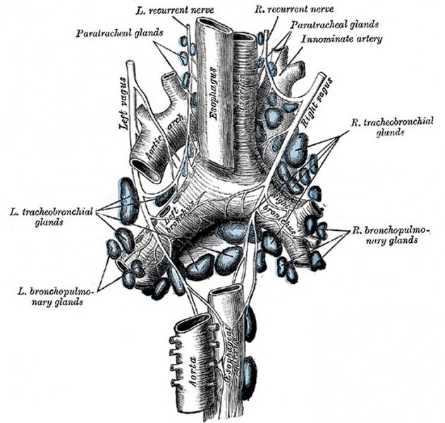
Tracheobronchial Lymph Glands
(From a figure designed by M. Hallé.)
Tracheobronchial Glands form four main groups:
- tracheal, on either side of the trachea
- bronchial, in the angles between the lower part of the trachea and bronchi and in the angle between the two bronchi
- bronchopulmonary, in the hilus of each lung
- pulmonary, in the lung substance, on the larger branches of the bronchi.
The afferents of the tracheobronchial glands drain the lungs and bronchi, the thoracic part of the trachea and the heart; some of the efferents of the posterior mediastinal glands also end in this group. Their efferent vessels ascend upon the trachea and unite with efferents of the internal mammary and anterior mediastinal glands to form the right and left bronchomediastinal trunks. The right bronchomediastinal trunk may join the right lymphatic duct, and the left the thoracic duct, but more frequently they open independently of these ducts into the junction of the internal jugular and subclavian veins of their own side.
In all town dwellers there are continually being swept into these glands from the bronchi and alveoli large quantities of the dust and black carbonaceous pigment that are so freely inhaled in cities. At first the glands are moderately enlarged, firm, inky black, and gritty on section; later they enlarge still further, often becoming fibrous from the irritation set up by the minute foreign bodies with which they are crammed, and may break down into a soft slimy mass or may calcify.
(Text from Gray's Anatomy 1918)
Gray's Lymphatic Anatomy: 592 Primary lymph sacs | 593 Lymph capillaries of the human conjunctiva | 594 Lymph capillaries from the human scrotum | 595 Lymph capillaries of the sole of the human foot | 596 Section through human tongue | 597 Lymph gland (Node) | 598 Lymph gland tissue | 599 Thoracic and right lymphatic ducts | 600 Modes of origin of thoracic duct | 601 Terminal collecting trunks of right side | 602 Lymph glands of the head | 603 Lymphatics of pharynx | 604 Lymphatics of the face | 605 Lymphatics of the Tongue | 606 Lymph glands of the upper extremity | 607 Lymphatics of the mamma | 608 Lymphatic vessels of the dorsal hand surface | 609 Lymph glands of popliteal fossa | 610 Superficial lymph glands and vessels of the lower extremity | 611 Parietal lymph glands of the pelvis | 612 Iliopelvic glands | 613 Lymphatics of stomach | 614 Lymphatics of stomach | 615 Lymphatics of cecum and vermiform process | 616 Lymphatics of cecum and vermiform process | 617 Lymphatics of Colon | 618 Lymphatic of the Bladder | 619 Lymphatics of the Prostate | 620 Lymphatics of the Uterus | 621 Lymphatics of the thorax and abdomen | 622 Tracheobronchial Lymph Glands | Gray's Anatomy | Historic Disclaimer | Lymphatic Development
- Gray's Images: Development | Lymphatic | Neural | Vision | Hearing | Somatosensory | Integumentary | Respiratory | Gastrointestinal | Urogenital | Endocrine | Surface Anatomy | iBook | Historic Disclaimer
| Historic Disclaimer - information about historic embryology pages |
|---|
 |
| iBook - Gray's Embryology | |
|---|---|

|
|
Reference
Gray H. Anatomy of the human body. (1918) Philadelphia: Lea & Febiger.
Cite this page: Hill, M.A. (2024, April 19) Embryology Gray0622.jpg. Retrieved from https://embryology.med.unsw.edu.au/embryology/index.php/File:Gray0622.jpg
- © Dr Mark Hill 2024, UNSW Embryology ISBN: 978 0 7334 2609 4 - UNSW CRICOS Provider Code No. 00098G
File history
Click on a date/time to view the file as it appeared at that time.
| Date/Time | Thumbnail | Dimensions | User | Comment | |
|---|---|---|---|---|---|
| current | 00:33, 15 February 2013 | | 737 × 700 (110 KB) | Z8600021 (talk | contribs) | The tracheobronchial lymph glands. (From a figure designed by M. Hallé.) (Text from Gray's Anatomy 1918) {{Gray Anatomy}} Category:Immune Category:Respiratory |
You cannot overwrite this file.
File usage
The following 3 pages use this file: