File:Gray0592.jpg
Gray0592.jpg (600 × 590 pixels, file size: 55 KB, MIME type: image/jpeg)
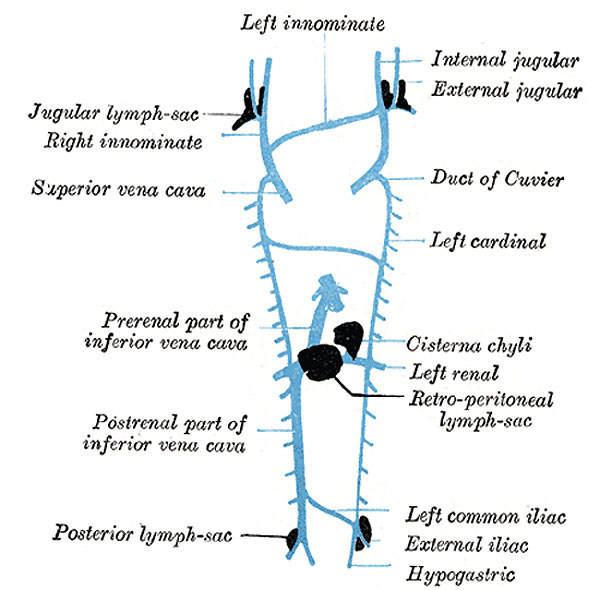
Primary Lymph Sacs
Scheme showing relative positions of primary lymph sacs based on the description given by Florence Sabin.
Development of the Lymphatic Vessels
The lymphatic system begins as a series of sacs 108 at the points of junction of certain of the embryonic veins. These lymph-sacs are developed by the confluence of numerous venous capillaries, which at first lose their connections with the venous system, but subsequently, on the formation of the sacs, regain them. The lymphatic system is therefore developmentally an offshoot of the venous system, and the lining walls of its vessels are always endothelial.
In the human embryo the lymph sacs from which the lymphatic vessels are derived are six in number; two paired, the jugular and the posterior lymph-sacs; and two unpaired, the retroperitoneal and the cisterna chyli. In lower mammals an additional pair, subclavian, is present, but in the human embryo these are merely extensions of the jugular sacs.
The position of the sacs is as follows: (1) jugular sac, the first to appear, at the junction of the subclavian vein with the primitive jugular; (2) posterior sac, at the junction of the iliac vein with the cardinal; (3) retroperitoneal, in the root of the mesentery near the suprarenal glands; (4) cisterna chyli, opposite the third and fourth lumbar vertebræ (Fig. 592). From the lymph-sacs the lymphatic vessels bud out along fixed lines corresponding more or less closely to the course of the embryonic bloodvessels. Both in the body-wall and in the wall of the intestine, the deeper plexuses are the first to be developed; by continued growth of these the vessels in the superficial layers are gradually formed. The thoracic duct is probably formed from anastomosing outgrowths from the jugular sac and cisterna chyli. At its connection with the cisterna chyli it is at first double, but the two vessels soon join.
All the lymph-sacs except the cisterna chyli are, at a later stage, divided up by slender connective tissue bridges and transformed into groups of lymph glands. The lower portion of the cisterna chyli is similarly converted, but its upper portion remains as the adult cisterna.
(Text from Gray's Anatomy 1918)
Gray's Lymphatic Anatomy: 592 Primary lymph sacs | 593 Lymph capillaries of the human conjunctiva | 594 Lymph capillaries from the human scrotum | 595 Lymph capillaries of the sole of the human foot | 596 Section through human tongue | 597 Lymph gland (Node) | 598 Lymph gland tissue | 599 Thoracic and right lymphatic ducts | 600 Modes of origin of thoracic duct | 601 Terminal collecting trunks of right side | 602 Lymph glands of the head | 603 Lymphatics of pharynx | 604 Lymphatics of the face | 605 Lymphatics of the Tongue | 606 Lymph glands of the upper extremity | 607 Lymphatics of the mamma | 608 Lymphatic vessels of the dorsal hand surface | 609 Lymph glands of popliteal fossa | 610 Superficial lymph glands and vessels of the lower extremity | 611 Parietal lymph glands of the pelvis | 612 Iliopelvic glands | 613 Lymphatics of stomach | 614 Lymphatics of stomach | 615 Lymphatics of cecum and vermiform process | 616 Lymphatics of cecum and vermiform process | 617 Lymphatics of Colon | 618 Lymphatic of the Bladder | 619 Lymphatics of the Prostate | 620 Lymphatics of the Uterus | 621 Lymphatics of the thorax and abdomen | 622 Tracheobronchial Lymph Glands | Gray's Anatomy | Historic Disclaimer | Lymphatic Development
- Gray's Images: Development | Lymphatic | Neural | Vision | Hearing | Somatosensory | Integumentary | Respiratory | Gastrointestinal | Urogenital | Endocrine | Surface Anatomy | iBook | Historic Disclaimer
| Historic Disclaimer - information about historic embryology pages |
|---|
 |
| iBook - Gray's Embryology | |
|---|---|

|
|
Reference
Gray H. Anatomy of the human body. (1918) Philadelphia: Lea & Febiger.
Cite this page: Hill, M.A. (2024, April 20) Embryology Gray0592.jpg. Retrieved from https://embryology.med.unsw.edu.au/embryology/index.php/File:Gray0592.jpg
- © Dr Mark Hill 2024, UNSW Embryology ISBN: 978 0 7334 2609 4 - UNSW CRICOS Provider Code No. 00098G
File history
Click on a date/time to view the file as it appeared at that time.
| Date/Time | Thumbnail | Dimensions | User | Comment | |
|---|---|---|---|---|---|
| current | 15:06, 14 February 2013 | | 600 × 590 (55 KB) | Z8600021 (talk | contribs) | ==Primary Lymph Sacs== Scheme showing relative positions of primary lymph sacs based on the description given by Florence Sabin. {{Gray Anatomy}} Category:Immune |
You cannot overwrite this file.
File usage
The following 3 pages use this file: