File:Gray0176.jpg
Gray0176.jpg (600 × 402 pixels, file size: 58 KB, MIME type: image/jpeg)
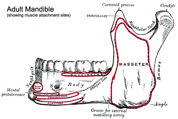
Adult Mandible
Adult mandible external view showing muscle attachment sites.
Surfaces
The lateral surface (Fig. 176) is flat and marked by oblique ridges at its lower part; it gives attachment throughout nearly the whole of its extent to the Masseter.
The external surface (Fig. 176) is marked in the median line by a faint ridge, indicating the symphysis or line of junction of the two pieces of which the bone is composed at an early period of life. This ridge divides below and encloses a triangular eminence, the mental protuberance, the base of which is depressed in the center but raised on either side to form the mental tubercle. On either side of the symphysis, just below the incisor teeth, is a depression, the incisive fossa, which gives origin to the Mentalis and a small portion of the Orbicularis oris. Below the second premolar tooth, on either side, midway between the upper and lower borders of the body, is the mental foramen, for the passage of the mental vessels and nerve. Running backward and upward from each mental tubercle is a faint ridge, the oblique line, which is continuous with the anterior border of the ramus; it affords attachment to the Quadratus labii inferioris and Triangularis; the Platysma is attached below it.
- Mandible Development: Week 8 outer view | Week 8 inner view | Week 12 outer view | Week 12 inner view | Week 12 Head outer view | Week 12 Head inner view | Birth | Childhood | Adult | Old Age | Small Animation | Large Animation | Muscle Attachments | Mandible Ossification | 1909 Mandible | embryo 18 mm | embryo 24 mm | embryo 28 mm | fetus 43 mm | fetus 65 mm | fetus 55 mm | fetus 95 mm | human 18-24-95 mm | Skull Development | Head Development
- Gray's Images: Development | Lymphatic | Neural | Vision | Hearing | Somatosensory | Integumentary | Respiratory | Gastrointestinal | Urogenital | Endocrine | Surface Anatomy | iBook | Historic Disclaimer
| Historic Disclaimer - information about historic embryology pages |
|---|
 |
| iBook - Gray's Embryology | |
|---|---|

|
|
Reference
Gray H. Anatomy of the human body. (1918) Philadelphia: Lea & Febiger.
Cite this page: Hill, M.A. (2024, April 23) Embryology Gray0176.jpg. Retrieved from https://embryology.med.unsw.edu.au/embryology/index.php/File:Gray0176.jpg
- © Dr Mark Hill 2024, UNSW Embryology ISBN: 978 0 7334 2609 4 - UNSW CRICOS Provider Code No. 00098G
File history
Click on a date/time to view the file as it appeared at that time.
| Date/Time | Thumbnail | Dimensions | User | Comment | |
|---|---|---|---|---|---|
| current | 17:41, 29 August 2011 | | 600 × 402 (58 KB) | S8600021 (talk | contribs) | |
| 17:40, 29 August 2011 | 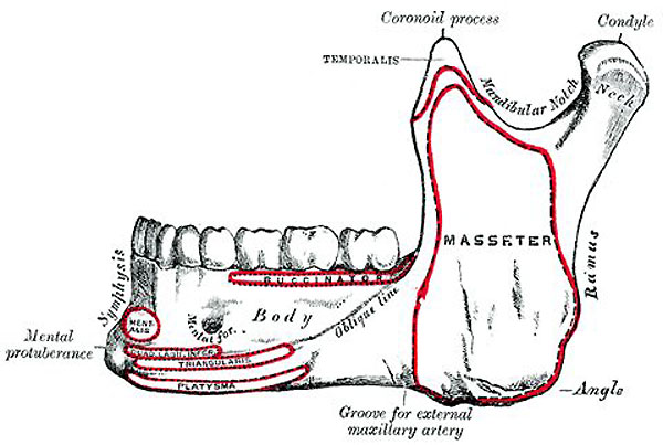 | 600 × 403 (52 KB) | S8600021 (talk | contribs) |
You cannot overwrite this file.
File usage
The following file is a duplicate of this file (more details):
The following 4 pages use this file: