File:Gray0119.jpg
Gray0119.jpg (450 × 307 pixels, file size: 22 KB, MIME type: image/jpeg)
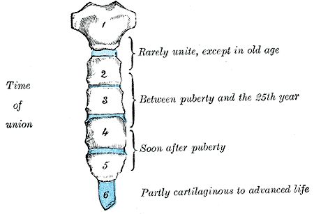
Fig. 119. Ossification of the Sternum
Fig. 118 - The sternum originally consists of two cartilaginous bars, situated one on either side of the median plane and connected with the cartilages of the upper nine ribs of its own side. These two bars fuse with each other along the middle line to form the cartilaginous sternum which is ossified from six centers: one for the manubrium, four for the body, and one for the xiphoid process.
Fig. 119 - Union of the various centers of the body begins about puberty, and proceeds from below upward; by the age of twenty-five they are all united. The xiphoid process may become joined to the body before the age of thirty, but this occurs more frequently after forty; on the other hand, it sometimes remains ununited in old age. In advanced life the manubrium is occasionally joined to the body by bone. When this takes place, however, the bony tissue is generally only superficial, the central portion of the intervening cartilage remaining unossified.
Fig. 120 - The ossific centers appear in the intervals between the articular depressions for the costal cartilages, in the following order: in the manubrium and first piece of the body, during the sixth month; in the second and third pieces of the body, during the seventh month of fetal life; in its fourth piece, during the first year after birth; and in the xiphoid process, between the fifth and eighteenth years. The centers make their appearance at the upper parts of the segments, and proceed gradually downward. 20 To these may be added the occasional existence of two small episternal centers, which make their appearance one on either side of the jugular notch; they are probably vestiges of the episternal bone of the monotremata and lizards. Occasionally some of the segments are formed from more than one center, the number and position of which vary.
Fig. 121 - Thus, the first piece may have two, three, or even six centers. When two are present, they are generally situated one above the other, the upper being the larger; the second piece has seldom more than one; the third, fourth, and fifth pieces are often formed from two centers placed laterally, the irregular union of which explains the rare occurrence of the sternal foramen, or of the vertical fissure which occasionally intersects this part of the bone constituting the malformation known as fissura sterni; these conditions are further explained by the manner in which the cartilaginous sternum is formed. More rarely still the upper end of the sternum may be divided by a fissure.
- Gray's Images: Development | Lymphatic | Neural | Vision | Hearing | Somatosensory | Integumentary | Respiratory | Gastrointestinal | Urogenital | Endocrine | Surface Anatomy | iBook | Historic Disclaimer
| Historic Disclaimer - information about historic embryology pages |
|---|
 |
| iBook - Gray's Embryology | |
|---|---|

|
|
Reference
Gray H. Anatomy of the human body. (1918) Philadelphia: Lea & Febiger.
Cite this page: Hill, M.A. (2024, April 20) Embryology Gray0119.jpg. Retrieved from https://embryology.med.unsw.edu.au/embryology/index.php/File:Gray0119.jpg
- © Dr Mark Hill 2024, UNSW Embryology ISBN: 978 0 7334 2609 4 - UNSW CRICOS Provider Code No. 00098G
File history
Click on a date/time to view the file as it appeared at that time.
| Date/Time | Thumbnail | Dimensions | User | Comment | |
|---|---|---|---|---|---|
| current | 15:24, 13 September 2009 | | 450 × 307 (22 KB) | S8600021 (talk | contribs) | Ossification of the sternum |
You cannot overwrite this file.
File usage
The following 4 pages use this file: