File:Gray0015.jpg

Original file (800 × 682 pixels, file size: 111 KB, MIME type: image/jpeg)
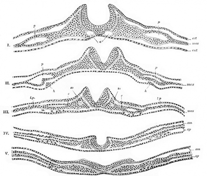
Neural Groove
A series of transverse sections through an embryo of the dog. (After Bonnet.)
The series shows the uprising of the neural folds to form the neural canal.
Section I is the most anterior. In V the neural plate is spread out nearly flat.
Section III, IV, and V the scattered cells represented between the entoderm and splanchnic layer of mesoderm are the vasoformative cells which give origin in front, according to Bonnet, to the heart tubes, h; l.p.
Lateral plate still undivided in I, II, and III; in IV and V split into somatic (sm) and splanchnic (sp) layers of mesoderm.
Legend
- a - Aortæ.
- c - Intermediate cell mass.
- ect. - Ectoderm.
- ent. - Entoderm. (endoderm)
- h - Rudiments of endothelial heart tubes.
- mes. - Mesoderm.
- p - Pericardium.
- so. - Primitive segment. (somite)
- Gray's Images: Development | Lymphatic | Neural | Vision | Hearing | Somatosensory | Integumentary | Respiratory | Gastrointestinal | Urogenital | Endocrine | Surface Anatomy | iBook | Historic Disclaimer
| Historic Disclaimer - information about historic embryology pages |
|---|
 |
| iBook - Gray's Embryology | |
|---|---|

|
|
Reference
Gray H. Anatomy of the human body. (1918) Philadelphia: Lea & Febiger.
Cite this page: Hill, M.A. (2024, April 24) Embryology Gray0015.jpg. Retrieved from https://embryology.med.unsw.edu.au/embryology/index.php/File:Gray0015.jpg
- © Dr Mark Hill 2024, UNSW Embryology ISBN: 978 0 7334 2609 4 - UNSW CRICOS Provider Code No. 00098G
File history
Click on a date/time to view the file as it appeared at that time.
| Date/Time | Thumbnail | Dimensions | User | Comment | |
|---|---|---|---|---|---|
| current | 13:45, 23 October 2010 | | 800 × 682 (111 KB) | S8600021 (talk | contribs) | ==Neural Groove== A series of transverse sections through an embryo of the dog. (After Bonnet.) The series shows the uprising of the neural folds to form the neural canal. Section I is the most anterior. In V the neural plate is spread out nearly fla |
You cannot overwrite this file.
File usage
The following 2 pages use this file: