File:Foster029.jpg
Original file (950 × 437 pixels, file size: 51 KB, MIME type: image/jpeg)
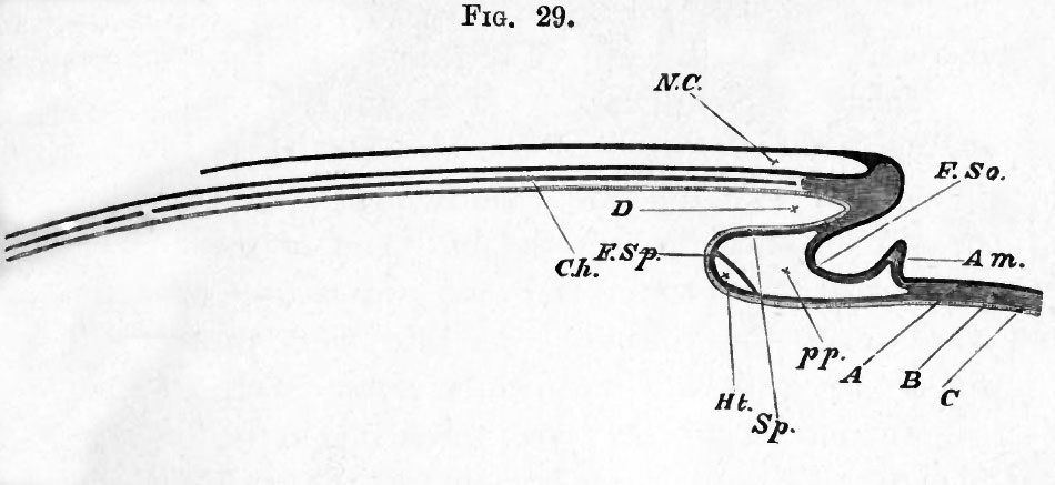
FIG. 29. DIAGRAMMATIC LONGITUDINAL SECTION THROUGH THE Axis OF AN EMBRYO.
The section is supposed to be made at a time when the headfold has commenced but the tail-fold has not yet appeared.
N.C. neural canal, closed in front but as yet open behind. Ch. notochord.
The section being taken in the middle line, the protovertebrae are of course not shewn.
In front of the notochord is seen a mass of uncleft mesoblast, which will eventually form part of the skull.
D. the commencing foregut or front part of the alimentary canal.
F. So. Somatopleure, raised up in its peripheral portion into the amniotic fold Am. Sp. Splanchnopleure. At Sp. it forms the under wall of the foregut ; at F. Sp. it is turning round and about to run forward.
Just at its turning point the cavity of the heart Ht. is being developed in its mesoblast. pp. pleuroperitoneal cavity. A epiblast, B mesoblast, C hypoblast, indicated in the rest of the figure by differences in the shading.
At the part where these three lines of reference end the mesoblast is as yet uncleft.
| Historic Disclaimer - information about historic embryology pages |
|---|
 |
Reference
Foster, M., Balfour, F. M., Sedgwick, A., & Heape, W. (1883). The Elements of Embryology. (2nd ed.). London: Macmillan and Co.
The Elements of Embryology (1883)
File history
Click on a date/time to view the file as it appeared at that time.
| Date/Time | Thumbnail | Dimensions | User | Comment | |
|---|---|---|---|---|---|
| current | 07:51, 9 January 2011 | | 950 × 437 (51 KB) | S8600021 (talk | contribs) | FIG. 29. DIAGRAMMATIC LONGITUDINAL SECTION THROUGH THE Axis OF AN EMBRYO. The section is supposed to be made at a time when the headfold has commenced but the tail-fold has not yet appeared. N.C. neural canal, closed in front but as yet open behind. Ch |
You cannot overwrite this file.
File usage
The following 3 pages use this file: