File:Foster028.jpg
Original file (629 × 848 pixels, file size: 77 KB, MIME type: image/jpeg)
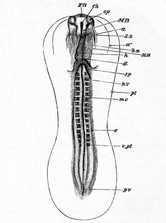
FIG. 28. AN EMBRYO CHICK OF ABOUT THIRTY-SIX HOURS, VIEWED FROM BELOW AS A TRANSPARENT OBJECT.
FB. the fore-brain or first cerebral vesicle, projecting from the sides of which are seen the optic vesicles, op. A definite head is now constituted, the backward limit of the somatopleure fold being indicated by the faint line S.O. Around the head are seen the two limbs of the amniotic head-fold : one, the true amnion a, closely enveloping the head, the other, the false amnion a', at some distance from it. The head is seen to project beyond the anterior limit of the pellucid area.
The splanchnopleure folds extend as far back as sp. Along its diverging limbs are seen the conspicuous venous roots of the vitelline veins, uniting to form the heart h, already established by the coalescence of two lateral halves which, continuing forward as the bulbus arteriosus b. a., is lost in the substance of the head just in front of the somatopleure fold.
HE. hind-brain ; ME. mid-brain ; p.v. and v.pl. mesoblastic somites ; ch. front end of notochord ; me. posterior part of notochord ; e. parietal mesoblast ; pi. outline of area pellucida ; pv. primitive streak.
| Historic Disclaimer - information about historic embryology pages |
|---|
 |
Reference
Foster, M., Balfour, F. M., Sedgwick, A., & Heape, W. (1883). The Elements of Embryology. (2nd ed.). London: Macmillan and Co.
The Elements of Embryology (1883)
File history
Click on a date/time to view the file as it appeared at that time.
| Date/Time | Thumbnail | Dimensions | User | Comment | |
|---|---|---|---|---|---|
| current | 07:43, 9 January 2011 | | 629 × 848 (77 KB) | S8600021 (talk | contribs) | FIG. 28. AN EMBRYO CHICK OF ABOUT THIRTY-SIX HOURS, VIEWED FROM BELOW AS A TRANSPARENT OBJECT. FB. the fore-brain or first cerebral vesicle, projecting from the sides of which are seen the optic vesicles, op. A definite head is now constituted, the bac |
You cannot overwrite this file.
File usage
The following 3 pages use this file: