File:Foster027.jpg
Original file (740 × 887 pixels, file size: 86 KB, MIME type: image/jpeg)
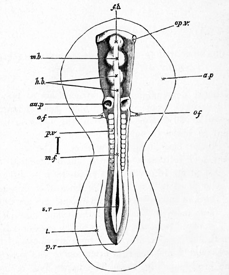
FIG. 27. EMBRYO OF THE CHICK BETWEEN THIRTY AND THIRTY-SIX HOURS, VIEWED FROM ABOVE AS AN OPAQUE OBJECT.
(Chromic acid preparation.)
f.b. front-brain : mb. mid-brain ; h. b. hind-brain ; op.v. optic vesicle ; au.p. auditory pit ; o.f. vitelline vein ; p.v. mesoblastic somite; m.f. line of junction of the medullary folds above the medullary canal ; s.r. sinus rhomboidalis ; t. tail-fold ; p.r. remains of primitive groove (not satisfactorily represented) ; a.p. area pellucida.
The line to the side between p.v. and m.f. represents the true length of the embryo.
The fiddle-shaped outline indicates the margin of the pellucid area. The head, which reaches as far back as o.f., is distinctly marked off; but neither the somatopleuric nor splanchnopleuric folds are shewn in the figure ; the latter diverge at the level of o./., the former considerably nearer the front, somewhere between the lines m.b. and h. b. The optic vesicles op.v. are seen bulging out beneath the superficial epiblast. The heart lying underneath the opaque body cannot be seen. The tail-fold t. is just indicated ; no distinct lateral folds are as yet visible in the region midway between head and tail. At m.f. the line of junction between the medullary folds is K still visible, being lost forwards over the cerebral vesicles, while behind may be seen the remains of the sinus rhomboidalis, s.r.
| Historic Disclaimer - information about historic embryology pages |
|---|
 |
Reference
Foster, M., Balfour, F. M., Sedgwick, A., & Heape, W. (1883). The Elements of Embryology. (2nd ed.). London: Macmillan and Co.
The Elements of Embryology (1883)
File history
Click on a date/time to view the file as it appeared at that time.
| Date/Time | Thumbnail | Dimensions | User | Comment | |
|---|---|---|---|---|---|
| current | 07:40, 9 January 2011 | | 740 × 887 (86 KB) | S8600021 (talk | contribs) | FIG. 27. EMBRYO OF THE CHICK BETWEEN THIRTY AND THIRTY-SIX HOURS, VIEWED FROM ABOVE AS AN OPAQUE OBJECT. (Chromic acid preparation.) f.b. front-brain : mb. mid-brain ; h. b. hind-brain ; op.v. optic vesicle ; au.p. auditory pit ; o.f. vitelline vein ; p |
You cannot overwrite this file.
File usage
The following 3 pages use this file: