File:Finley1923 fig09.jpg
From Embryology

Size of this preview: 342 × 600 pixels. Other resolution: 456 × 800 pixels.
Original file (456 × 800 pixels, file size: 49 KB, MIME type: image/jpeg)
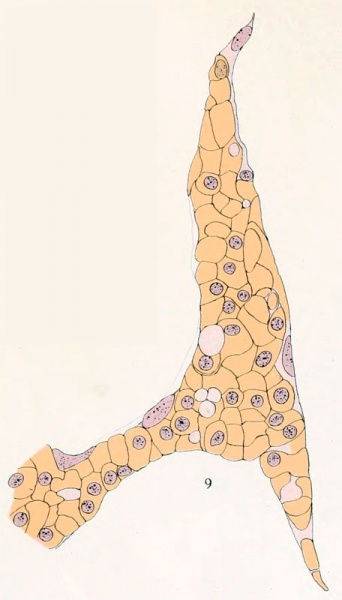
Figure 9
Drawing of a growing tip at the edge of the head plexus in a human embryo 28 mm. in length (No. 1240a, total mount). Two cells with clear, colorless cytoplasm may be observed. X930.
- 1923 Head Subcutaneous Plexus: Plate 1 | Plate 2 | Fig 1 | Fig 2 | Fig 3 | Fig 4 | Fig 5 | Fig 6 | Fig 7 | Fig 8 | Fig 9 | Fig 10 | Fig 11 | Fig 12 | Fig 13 | Carnegie No.71 | Historic Disclaimer
| Historic Disclaimer - information about historic embryology pages |
|---|
 |
Reference
Finley EB. The development of the subcutaneous vascular plexus in the head of the human embryo. (1923) Contributions to Embryology Carnegie Institution No. 71: 155-161.
Cite this page: Hill, M.A. (2024, April 19) Embryology Finley1923 fig09.jpg. Retrieved from https://embryology.med.unsw.edu.au/embryology/index.php/File:Finley1923_fig09.jpg
- © Dr Mark Hill 2024, UNSW Embryology ISBN: 978 0 7334 2609 4 - UNSW CRICOS Provider Code No. 00098G
File history
Click on a date/time to view the file as it appeared at that time.
| Date/Time | Thumbnail | Dimensions | User | Comment | |
|---|---|---|---|---|---|
| current | 09:35, 14 August 2012 | | 456 × 800 (49 KB) | Z8600021 (talk | contribs) | ==Figure 9== {{Finley1923}} |
You cannot overwrite this file.